
Pediatric & Congenital Cardiology
Tetralogy of Fallot: A Medical-Legal Guide for Attorneys
- Published
- November 10, 2026
- Last Reviewed
- November 10, 2026
- Author
- Asif Masood, MD, MSc
- Reading Time
- 14 min
- Category
- Pediatric & Congenital Cardiology
Tetralogy of Fallot (TOF) is the most common cyanotic congenital heart defect. It occurs in about 1 in 3,600 births and makes up 7–10% of all congenital heart malformations (Silva et al., Pediatric Cardiology, 2020). Modern surgery has made survival into adulthood the expected outcome. That success means TOF can come up in litigation at any stage of life: a newborn diagnosed late, an operation in infancy, a missed sign of a "tet spell," or problems decades after repair when surveillance falls short. To evaluate these cases, attorneys need to understand three things. TOF is a spectrum, not one disease. Several treatment strategies are accepted. And many late complications are part of the condition's expected course after repair, not evidence of an error. This article is for education only. It is not legal advice, and it offers no opinion on liability in any specific case.
Hypothetical Scenario (Fictional, for Educational Purposes Only)
Baby M. is born at term at a community hospital. A routine prenatal anatomy scan was read as normal. In the nursery, a soft systolic murmur is noted on day 1. The newborn pulse oximetry screen at 26 hours shows 96% in the right hand and 95% in the foot, and the screen is recorded as a pass. The murmur is documented as "likely a flow murmur; recheck at follow-up," and Baby M. goes home on day 2.
At the 1-week visit, the murmur is louder. The pediatrician notes "good color" and plans a recheck at 1 month. At 6 weeks, the parents report brief episodes of turning "dusky" while crying, which resolve on their own. Office oxygen saturation is 88%. The child is referred to cardiology, with an appointment three weeks out.
At 8 weeks, Baby M. has a prolonged episode of deep cyanosis and limpness after a long crying spell and is taken to the emergency department. Saturation is 55%. Echocardiography confirms TOF with severe infundibular (muscular outflow) obstruction. The child is stabilized and undergoes complete repair at 10 weeks. Afterward, there are concerns about a possible hypoxic brain injury.
This scenario raises several questions an expert would consider. Was a murmur on day 1 with a passed screen enough reason to get an echocardiogram before discharge? Were the parents' reports of dusky episodes recognized as possible hypercyanotic spells? Was a three-week wait for cardiology reasonable once saturation was 88% and spells were suspected? And did any delay cause the neurological outcome, or would the severe spell have happened anyway? The scenario is built to raise these questions, not to answer them.
Understanding the Anatomy
TOF results from forward displacement of the outlet septum during fetal development. That single developmental error produces four linked features:
- Right ventricular outflow tract obstruction (RVOTO). Narrowing below, at, or above the pulmonary valve limits blood flow to the lungs.
- Ventricular septal defect (VSD). A large hole between the two pumping chambers.
- Overriding aorta. The aorta sits over the VSD and receives blood from both ventricles.
- Right ventricular hypertrophy. The right ventricle thickens because it pumps against the obstruction.
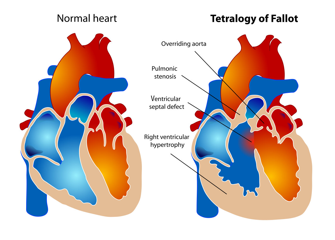
The severity of the outflow obstruction drives the clinical picture. Mild obstruction may cause little cyanosis (a "pink tet"). Severe obstruction or pulmonary atresia makes lung blood flow depend on the ductus arteriosus. These babies need prostaglandin E1 to keep the duct open, and they need early intervention (Silva et al., 2020; Bashir et al., Pediatric Cardiology, 2026). The obstruction often worsens over the first months of life.
About 20% of patients have a genetic syndrome, most commonly 22q11.2 deletion or trisomy 21. These syndromes are linked to more illness, higher mortality, and longer hospital stays, independent of the quality of care (Silva et al., 2020). A right aortic arch appears in 15–25% of patients. An abnormal coronary artery crossing the outflow tract appears in 3–5%, which matters for surgical planning (Dodge-Khatami et al., Pediatric Cardiac Surgery, 5th ed., 2023).
Hypercyanotic ("Tet") Spells
Tet spells are episodes of sudden, severe drop in oxygen. They are often triggered by crying, fever, dehydration, a warm bath, or exertion: anything that lowers systemic vascular resistance or increases the outflow spasm (Dodge-Khatami et al., 2023). During a spell, the murmur may soften or disappear because little blood crosses the narrowed outflow tract. A quieter murmur can therefore mean the child is getting worse, not better.
Prolonged spells can cause hypoxic brain injury, seizures, stroke, or death. In standard practice, an unrepaired infant who has spells needs urgent cardiology evaluation, and intervention is usually brought forward. Parents' descriptions of turning blue, dusky, or limp episodes are often the first sign. How those reports were documented and acted on is often central to litigation.
Prenatal Diagnosis
Fetal echocardiography diagnoses TOF with a high degree of accuracy. However, it cannot fully predict how severe the pulmonary stenosis will be after birth (Dodge-Khatami et al., 2023). The four-chamber view used in routine anatomy scans may look normal in TOF, because the defect affects the outflow tracts. Detection depends on whether the outflow tract views were obtained and how they were interpreted.
When reviewing an alleged missed prenatal diagnosis, experts usually consider gestational age, maternal body habitus, fetal position, image quality, whether outflow views were recorded, and the protocol in place at that time and place. A missed prenatal diagnosis does not, by itself, establish a deviation from accepted practice. Its effect on outcome also differs: many infants with TOF are stable at birth and gain little from prenatal diagnosis, while ductal-dependent infants may gain substantially.
Postnatal Recognition and Pulse Oximetry Screening
Critical congenital heart disease (CCHD) screening was added to the US Recommended Uniform Screening Panel in 2011 and was adopted in all states and territories by July 2018 (Oster et al., Pediatrics, 2025). TOF is one of the core target lesions of screening (Olsen and Puri, Pediatrics in Review, 2022).
Key points for attorneys:
- Visible cyanosis is unreliable. Cyanosis usually becomes visible only at saturations around 80% or below. It is especially hard to see in infants with darker skin (Wasserman et al., Journal of the American Society of Echocardiography, 2021).
- Screening misses cases. A 2018 Cochrane review of more than 400,000 newborns found an overall sensitivity of 76.3% and specificity of 99.9% for CCHD (Olsen and Puri, 2022). Many infants with TOF and mild obstruction have normal saturations at 24–48 hours and pass.
- The algorithm has changed. The original protocol was modified in 2018. Passing now requires at least 95% in both the right hand and the foot, and only one repeat screen is allowed for a borderline result. The AAP issued an updated algorithm and recommendations in 2025 (Olsen and Puri, 2022; Oster et al., 2025). The protocol in effect at the birth hospital on the date of birth is the relevant reference.
- Delayed diagnosis still happens. In a Massachusetts population study, 13.8% of CCHD cases were diagnosed after discharge home. TOF was among the most common defects diagnosed late. Delivery outside a tertiary hospital was associated with a 3.6-fold higher likelihood of delayed diagnosis (Liberman et al., Pediatrics, 2014).
A passed screen does not rule out TOF. A newborn murmur, especially a harsh systolic ejection murmur, is an independent finding that experts evaluate separately.

Diagnostic Evaluation
Echocardiography is the main diagnostic test in almost all cases. It defines the anatomy, measures the outflow gradient, and usually identifies the coronary artery pattern. Electrocardiograms show right ventricular hypertrophy and right axis deviation but are not diagnostic. Chest x-rays may show the classic "boot-shaped" heart with dark lung fields. Most patients no longer need cardiac catheterization before repair (Dodge-Khatami et al., 2023).
Treatment: Accepted Strategies
Asymptomatic or minimally symptomatic infants. The American Association for Thoracic Surgery (AATS) 2022 Expert Consensus states that elective complete repair between 3 and 6 months of age is reasonable (Class IIa). Some centers repair earlier with similarly good outcomes (Miller et al., Journal of Thoracic and Cardiovascular Surgery, 2023). Other sources describe elective repair across the first year, roughly 3–12 months (Silva et al., 2020; Bashir et al., 2026). Repair timing varies within an accepted range, and timing within that range is not, by itself, a deviation from accepted practice.
Symptomatic neonates. Newborns with severe cyanosis, ductal dependence, or spells need early intervention. There are two accepted pathways:
- Primary complete repair in the newborn period.
- Staged repair: initial palliation (a surgical shunt, ductal stent, or outflow tract stent) followed later by complete repair.
The AATS consensus states that in standard-risk symptomatic neonates, excellent results can be achieved with either approach. Institutional expertise partly drives the choice, and observational comparisons do not show substantial differences (Miller et al., 2023). A 2026 meta-analysis of 4,628 neonates confirmed that the best approach remains debated (Bashir et al., 2026). A claim built only on the choice of strategy therefore faces a substantial body of literature supporting both approaches.
Surgical considerations. Complete repair closes the VSD and relieves the outflow obstruction. When the pulmonary valve annulus is small, a transannular patch may be needed. The patch relieves obstruction but makes the pulmonary valve permanently leaky (pulmonary regurgitation). Valve-sparing repair is preferred when the anatomy allows it, but it is not always possible. Repair before 3 months has been associated with more frequent transannular patch use in some series (Miller et al., 2023).
Early Postoperative Complications
Recognized complications after TOF repair include low cardiac output, junctional ectopic tachycardia, heart block, residual VSD, residual outflow obstruction, bleeding, infection, and chylothorax. Many of these occur even with appropriate care. In litigation, the question is usually not whether a complication happened. It is whether the complication was recognized and managed in a timely, appropriate way: for example, whether a significant residual defect seen on intraoperative or postoperative echocardiography was addressed, or whether heart block was managed according to accepted pacing indications.
Long-Term Outcomes and Surveillance
Late problems after repair are expected, not exceptional. The American Heart Association states that right ventricular outflow dysfunction (stenosis, regurgitation, or both) is nearly universal after TOF repair. It drives rising illness and mortality with age (Geva et al., Circulation, 2024).
- Pulmonary regurgitation. Moderate-to-severe regurgitation develops in 40–85% of patients within 5–10 years of repair. It leads to progressive right ventricular dilation, dysfunction, exercise intolerance, and arrhythmia (Cohen et al., Journal of the American College of Cardiology, 2021).
- Pulmonary valve replacement (PVR). Most patients will eventually need surgical or transcatheter PVR. The 2025 ACC/AHA adult congenital heart disease guideline recommends PVR for symptomatic adults with moderate or greater pulmonary valve dysfunction. For asymptomatic adults, PVR is considered reasonable when at least two volume, function, or exercise criteria are met, such as a right ventricular end-systolic volume index above 80 mL/m², right ventricular ejection fraction of 46% or less, or a progressive decline in exercise capacity (Gurvitz et al., Journal of the American College of Cardiology, 2026). The 2026 STS/WSPCHS/ECHSA guidelines and consensus documents add child-specific guidance and emphasize symptoms and ventricular volumes (Nelson et al., Annals of Thoracic Surgery, 2026; Stephens et al., Annals of Thoracic Surgery, 2026). These thresholds have changed over time, so care must be judged against the guidance in effect when the decision was made.
- Arrhythmias and sudden death. Repaired TOF carries a risk of atrial flutter and fibrillation, sustained ventricular tachycardia, and sudden cardiac death. The classic rate of sudden death is about 1.5 per 1,000 patient-years. Risk is low in childhood and accelerates in the third and fourth decades of life (Gurvitz et al., 2026). An implantable defibrillator is reasonable for adults at high risk.
- Other issues. Residual VSD, tricuspid regurgitation, left ventricular dysfunction (up to 20% of adults), and aortic root dilation (Gurvitz et al., 2026).
- Neurodevelopment. Children with cyanotic heart disease are at risk for developmental, learning, and attention problems. Contributing factors include genetic syndromes, prenatal brain development, and surgical and perioperative factors, so it is often hard to attribute deficits to any single event.
Lifelong follow-up with a congenital cardiologist, including periodic imaging and rhythm assessment, is standard. Loss to follow-up during the shift from pediatric to adult care is a well-recognized gap. Late cases often focus on whether surveillance was arranged, whether imaging findings were acted on, and whether the patient was referred for PVR or electrophysiology evaluation.
Common Targets for Litigation
The table below summarizes allegations that come up often and the medical questions experts typically examine. None of these allegations automatically establishes a deviation from accepted practice.
Missed prenatal diagnosis
- Medical Questions Commonly Examined
- Were outflow tract views obtained? Image quality, gestational age, protocol at the time; would prenatal diagnosis have changed the outcome?
Discharge with an unexplained murmur
- Medical Questions Commonly Examined
- Character of the murmur; was echocardiography or cardiology input reasonably available; was follow-up timely?
Pulse oximetry error
- Medical Questions Commonly Examined
- Was the screen done, correctly timed, and interpreted with the algorithm in effect? Was a failed or borderline screen acted on?
Failure to recognize tet spells
- Medical Questions Commonly Examined
- Were parental reports documented? Was urgent evaluation arranged? Were families taught what to watch for?
Delayed cardiology referral or repair
- Medical Questions Commonly Examined
- Urgency of the referral relative to symptoms and saturation; was repair timing within the accepted range?
Choice of primary vs staged repair
- Medical Questions Commonly Examined
- Both strategies are accepted; was the choice reasonable given anatomy, risk factors, and institutional expertise?
Surgical or perioperative complications
- Medical Questions Commonly Examined
- Was the complication a recognized risk? Was it detected and managed promptly? Was informed consent adequate?
Failure of long-term surveillance
- Medical Questions Commonly Examined
- Was follow-up arranged? Were imaging and rhythm findings acted on? Was PVR or defibrillator evaluation timely?
Arrhythmic death in adulthood
- Medical Questions Commonly Examined
- Were known risk markers present and evaluated? Was care consistent with the guidance in effect at the time?
| Allegation | Medical Questions Commonly Examined |
|---|---|
| Missed prenatal diagnosis | Were outflow tract views obtained? Image quality, gestational age, protocol at the time; would prenatal diagnosis have changed the outcome? |
| Discharge with an unexplained murmur | Character of the murmur; was echocardiography or cardiology input reasonably available; was follow-up timely? |
| Pulse oximetry error | Was the screen done, correctly timed, and interpreted with the algorithm in effect? Was a failed or borderline screen acted on? |
| Failure to recognize tet spells | Were parental reports documented? Was urgent evaluation arranged? Were families taught what to watch for? |
| Delayed cardiology referral or repair | Urgency of the referral relative to symptoms and saturation; was repair timing within the accepted range? |
| Choice of primary vs staged repair | Both strategies are accepted; was the choice reasonable given anatomy, risk factors, and institutional expertise? |
| Surgical or perioperative complications | Was the complication a recognized risk? Was it detected and managed promptly? Was informed consent adequate? |
| Failure of long-term surveillance | Was follow-up arranged? Were imaging and rhythm findings acted on? Was PVR or defibrillator evaluation timely? |
| Arrhythmic death in adulthood | Were known risk markers present and evaluated? Was care consistent with the guidance in effect at the time? |
Medical Causation
Causation is often the most disputed issue in TOF cases. Experts commonly consider:
- Natural history. Outflow obstruction tends to progress, and spells can happen suddenly even in children under appropriate surveillance.
- Baseline risk. Genetic syndromes, prematurity, pulmonary atresia, and small pulmonary arteries independently affect survival and neurodevelopment.
- Timing. When was the condition reasonably detectable, and would earlier recognition have changed management or outcome? Many infants with TOF would have had the same elective repair whenever they were diagnosed.
- Mechanism of injury. Is there objective evidence (imaging, lab values, documented saturations) linking a specific hypoxic event to the claimed injury, as opposed to other factors that contribute to neurodevelopmental problems?
- Probability vs possibility. The question is whether earlier action would more likely than not have changed the outcome, not whether it might have.
Medical Records That Often Matter
- Prenatal ultrasound reports and stored images, especially outflow tract views
- Nursery records: murmur documentation, pulse oximetry values, screening times, discharge exam
- Pediatric office notes and telephone triage logs documenting parental concerns
- Referral records, including appointment dates and urgency designations
- Emergency department records, saturations, and transport records
- Echocardiograms (images, not just reports) and catheterization data
- Operative reports, intraoperative echocardiograms, and perfusion records
- ICU flowsheets, rhythm strips, and lab trends
- Long-term cardiology notes, MRI reports, Holter monitoring, exercise tests, and transition-of-care records
- Neuroimaging and developmental assessments
Common Misconceptions
- "The baby passed pulse oximetry, so the heart was normal." Screening misses a meaningful share of CCHD, and TOF with mild obstruction often passes.
- "A louder murmur is reassuring; a softer one is better." During a tet spell, the murmur may soften because flow across the narrowed outflow tract falls.
- "Repair cures TOF." Repair is palliative in a lifelong sense. Pulmonary regurgitation, reoperations, and arrhythmias are expected parts of the course.
- "A later operation means the first one was done wrong." Most patients with repaired TOF eventually need PVR or other procedures.
- "Neurodevelopmental problems must result from a medical error." Many factors contribute, including genetics and prenatal brain development.
The Value of Pediatric Cardiology Expertise
Evaluating TOF cases requires understanding fetal imaging, neonatal physiology, how screening programs work, the range of accepted surgical strategies, postoperative care, and decades of long-term outcome data. A pediatric and congenital cardiologist can put each decision in the context of the guidance and evidence available at the time, separate expected sequelae from potentially avoidable harm, and address causation with scientific rigor.
Key Takeaways for Attorneys
- TOF is a spectrum. Severity of outflow obstruction determines urgency.
- Visible cyanosis is an unreliable sign; saturation measurements matter.
- Pulse oximetry screening is valuable but imperfect, and TOF can pass.
- Parental reports of dusky or blue spells can signal hypercyanotic spells and warrant urgent attention.
- Elective repair between about 3 and 6 months is reasonable under AATS consensus, and timing varies across centers.
- Primary and staged repair are both accepted for symptomatic neonates.
- Pulmonary regurgitation and the eventual need for valve replacement are expected after repair.
- Late arrhythmia and sudden death risk rise in adulthood, making lifelong surveillance essential.
- Causation requires analysis of natural history, baseline risk, and timing, not just the existence of a delay.
- Care must be judged against the guidance and protocols in effect at the time it was given.
References
- Silva et al., Pediatric Cardiology, 2020.
- Bashir et al., Pediatric Cardiology, 2026.
- Dodge-Khatami et al., in Mavroudis, Backer, and Anderson, eds., Pediatric Cardiac Surgery, 5th ed., Wiley, 2023.
- Oster et al., Pediatrics, 2025.
- Olsen and Puri, Pediatrics in Review, 2022.
- Wasserman et al., Journal of the American Society of Echocardiography, 2021.
- Liberman et al., Pediatrics, 2014.
- Miller et al., Journal of Thoracic and Cardiovascular Surgery, 2023.
- Geva et al., Circulation, 2024.
- Cohen et al., Journal of the American College of Cardiology, 2021.
- Gurvitz et al., Journal of the American College of Cardiology, 2026.
- Nelson et al., Annals of Thoracic Surgery, 2026.
- Stephens et al., Annals of Thoracic Surgery, 2026.
Educational Disclaimer
This article is provided for educational purposes only and does not constitute legal or medical advice. Every case is unique and should be evaluated on its individual facts and medical records. Reading this article does not create an expert-client relationship.
About the Author
Asif Masood, MD, MSc is double board-certified in General Pediatrics and Pediatric Cardiology. Expert witness services in general pediatrics and pediatric & congenital cardiology, complemented by independent, non-testimony medical consulting across a broad range of medical specialties. Through The Verdict MD, he serves attorneys, law firms, insurers, and healthcare organizations nationwide.
Learn about Dr. Masood's pediatric cardiology expert witness services for independent review of the medical questions in your case.