
Pediatric & Congenital Cardiology
Coarctation of the Aorta: A Medical-Legal Guide for Attorneys
- Published
- November 12, 2026
- Last Reviewed
- November 12, 2026
- Author
- Asif Masood, MD, MSc
- Reading Time
- 19 min
- Category
- Pediatric & Congenital Cardiology
This is educational material for legal professionals. It is not legal advice and expresses no opinion on liability in any specific case.
Introduction
Coarctation of the aorta (CoA) is a narrowing of the aorta, the main artery carrying blood from the heart to the body. It is one of the congenital heart defects most often diagnosed late, frequently after a newborn has gone home from the nursery. It also comes up often in medical-legal review, for three reasons:
- It can be silent at birth and turn life-threatening days or weeks later.
- Routine screening tools (prenatal ultrasound and newborn pulse oximetry) miss a large share of cases.
- The physical examination, especially checking femoral (groin) pulses and comparing arm and leg blood pressures, still plays a central role in detection.
A population-based study in Pediatrics found that prenatal ultrasound and pulse oximetry both had low sensitivity for CoA, and that nearly half of newborns with isolated CoA left the hospital undiagnosed (Lannering et al., "Late Diagnosis of Coarctation Despite Prenatal Ultrasound and Postnatal Pulse Oximetry," Pediatrics, 2015). Understanding why requires understanding the underlying physiology.
Hypothetical Clinical Vignette (Fictional, for Educational Purposes Only)
A full-term baby boy is born after an uncomplicated pregnancy. The routine 20-week anatomy ultrasound was reported as normal. In the nursery, the newborn exam documents "normal heart sounds, no murmur." Femoral pulses are recorded with a template checkbox as "2+ bilaterally." At 26 hours of age, the pulse oximetry screen shows 98% in the right hand and 97% in the foot, a pass. He is discharged at 36 hours.
At the 3-day weight check, the mother says he is feeding more slowly and sweating with feeds. Weight is 8% below birth weight. The note says "breastfeeding difficulty; lactation referral." No pulse exam is documented.
On day 9, the mother calls the after-hours line because he is "breathing fast and won't eat." She is told to bring him in the next morning. On day 10 he arrives at the emergency department gray, with a heart rate of 210, a respiratory rate of 80, cool mottled legs, no palpable femoral pulses, an enlarged liver, and severe metabolic acidosis (lactate 12). An echocardiogram shows severe coarctation, a closed ductus arteriosus, and markedly reduced left ventricular function.
Prostaglandin E1 is started and he is transferred to a cardiac center. After stabilization he undergoes surgical repair. His course is complicated by acute kidney injury and necrotizing enterocolitis. He survives, with recovery of heart function.
Questions an expert reviewer might consider:
- Was the nursery pulse exam actually performed, or was it a template default?
- Did the passing pulse oximetry screen give false reassurance?
- Should feeding difficulty and sweating at day 3 have prompted a cardiac exam?
- Was the after-hours triage response appropriate?
- Would earlier diagnosis have changed the kidney and bowel injury, and with what degree of medical probability?
The vignette is intentionally mixed. Some findings may represent reasonable care, and some injuries might have occurred even with earlier recognition.
Understanding Coarctation of the Aorta
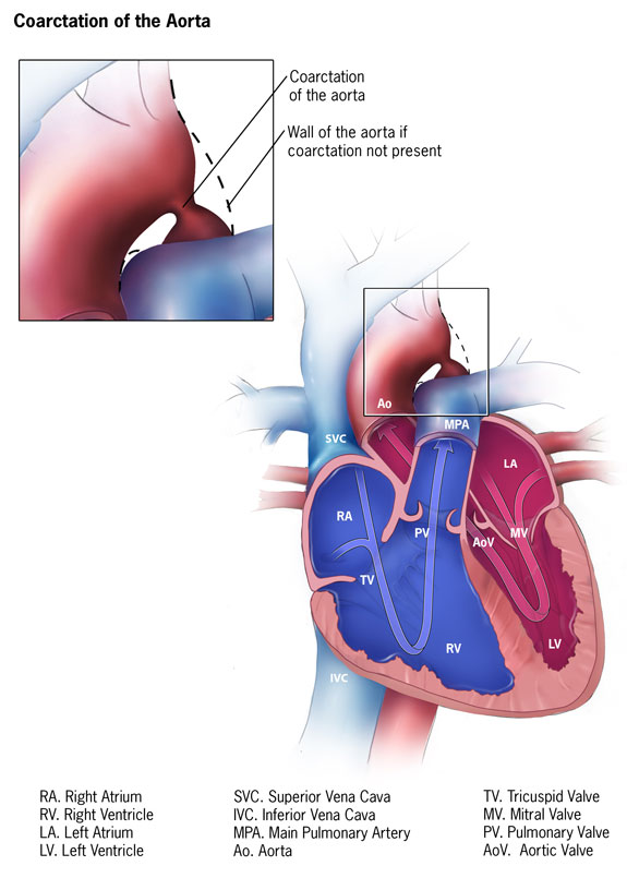
Anatomy. The narrowing usually occurs in the region where the ductus arteriosus joins the aorta, just past the artery to the left arm (the left subclavian artery) (Isselbacher et al., "2022 ACC/AHA Guideline for the Diagnosis and Management of Aortic Disease," Circulation/JACC, 2022). It may be a discrete "shelf" or a longer narrowing, sometimes with underdevelopment of the aortic arch (arch hypoplasia).
Associated conditions. CoA frequently occurs with other abnormalities:
- Bicuspid aortic valve (an aortic valve with two leaflets instead of three) in at least 50% of cases (Isselbacher et al., 2022), and more than 60% in the 2025 adult congenital heart disease guideline (Gurvitz et al., "2025 ACC/AHA/HRS/ISACHD/SCAI Guideline for the Management of Adults With Congenital Heart Disease," Journal of the American College of Cardiology, 2026).
- Ventricular septal defect, patent ductus arteriosus, and aortic arch hypoplasia (Dijkema et al., "Diagnosis, Imaging and Clinical Management of Aortic Coarctation," Heart, 2017).
- Complex congenital heart disease, and genetic conditions such as Turner syndrome (standard clinical knowledge).
The role of the ductus arteriosus. Before birth, the ductus arteriosus is a normal blood vessel connecting the pulmonary artery to the aorta. It normally closes in the first days of life. While it is open, it can carry blood around the narrowed segment to the lower body, masking the coarctation. As it closes, the obstruction can suddenly become severe.
Critical CoA typically appears after sudden constriction or closure of the ductus. A baby can pass pulse oximetry screening right after birth and then present several weeks later directly in cardiogenic shock, without many earlier warning signs (Hysko et al., "Advances in the Treatment of Neonatal Coarctation of the Aorta," Pediatrics, 2025).
Two clinical patterns.
- Critical neonatal coarctation: presents in the first days to weeks of life, often with heart failure or shock when the ductus closes.
- Later-presenting coarctation: less severe narrowing allows collateral vessels (natural bypass channels) to develop. The child or adult may have no symptoms and come to attention through a murmur, high blood pressure, or weak leg pulses (Backer et al., "Coarctation of the Aorta," Pediatric Cardiac Surgery, 5th ed., 2023).
Natural history if untreated. Critical neonatal CoA can progress rapidly to multiorgan failure, including kidney failure and necrotizing enterocolitis, and death (Hysko et al., 2025). In older patients, untreated CoA can be complicated by aortic dissection, heart failure, ruptured brain aneurysm, and complications of hypertension (Isselbacher et al., 2022).
Prenatal Diagnosis
CoA is one of the hardest heart defects to diagnose before birth. While the fetus is in the womb, the ductus is wide open and carries a large share of blood flow, so the aortic narrowing may not be apparent. Fetal findings are often indirect, such as a right-sided heart larger than the left or a small aortic arch, and these findings can also occur in normal fetuses.
Prenatal diagnosis remains difficult despite echocardiographic screening (Dijkema et al., 2017). The American Academy of Pediatrics (AAP) notes that prenatal detection of critical congenital heart disease is still under 60% in many areas of the United States, and lower detection has been linked to poverty, rural residence, and public insurance (Oster et al., "Newborn Screening for Critical Congenital Heart Disease: A New Algorithm and Other Updated Recommendations," Pediatrics, 2025). In one tertiary-center study, fetal diagnosis of CoA increased from 20.5% to 30.5% between two time periods (Geggel, "Coarctation of the Aorta: Delay in Diagnosis and Referral Basis From Infancy to Adulthood," Journal of Pediatrics, 2022).
Two practical points follow:
- A normal routine anatomy scan does not exclude CoA, and a missed prenatal diagnosis alone does not establish a deviation from accepted practice.
- Prenatal suspicion of CoA frequently produces false positives. Babies with suspected CoA are typically monitored after birth with serial echocardiograms as the ductus closes. Whether that monitoring plan was followed is often more relevant than whether the fetal scan was conclusive.
Postnatal Recognition
Symptoms. Early symptoms are often nonspecific: fast breathing, increased breathing effort, restlessness, irritability, excessive sweating, and poor feeding (Hysko et al., 2025). These overlap with common newborn problems, such as feeding difficulties, infection, and reflux. That overlap is central to many disputes about when CoA "should" have been recognized.
Physical examination. The key findings are:
- Weak, delayed, or absent femoral pulses
- Upper-to-lower-extremity systolic blood pressure difference greater than 20 mm Hg
- Pale or poorly perfused legs
- Sometimes differential cyanosis, meaning the legs are bluer than the arms because the ductus supplies them with low-oxygen blood (Hysko et al., 2025)
These findings have important limitations:
- Blood pressure differences can be hard to measure in awake newborns, and repeated measurements are needed.
- When heart function is severely reduced, or when a large VSD is present, the pressure difference may be minimal (Hysko et al., 2025).
- While the ductus is open, femoral pulses may feel normal.
Murmur. A murmur may be present from flow across the narrowing or an associated bicuspid valve (Dijkema et al., 2017), but many babies with critical CoA have no significant murmur. A "no murmur" entry does not exclude CoA.
Shock presentation. The newborn whose ductus closes presents in shock: fast breathing and heart rate, pallor, absent leg pulses, thready arm pulses, possible low blood pressure even in the arms, and an enlarged liver (Backer et al., 2023). This picture is easily mistaken for sepsis. Standard practice is to consider ductal-dependent heart disease, and start prostaglandin if it is suspected, in any newborn presenting with shock in the first weeks of life.
Later childhood and adulthood. CoA presents with high blood pressure in the arms, which is frequently attributed to kidney disease or "essential" hypertension. In the tertiary-center review, about 25% of patients were diagnosed after age 1. Decreased femoral pulses or hypertension were infrequently documented by referring physicians, and hypertension was typically attributed to a renal or essential cause (Geggel, 2022). The 2025 adult congenital guideline notes that CoA can be missed in childhood and first diagnosed in adulthood, usually during evaluation of resistant hypertension (Gurvitz et al., 2026).
Pulse Oximetry Screening
Background. Critical congenital heart disease (CCHD) screening was added to the US Recommended Uniform Screening Panel in 2011. All states and territories had adopted it by July 2018. The screening has reduced infant mortality and is cost-effective (Oster et al., 2025). It is designed to detect heart conditions that cause low blood oxygen.
Procedure. The AAP has advised screening after 24 hours of age, or shortly before discharge if earlier, in the right hand and either foot, followed by a diagnostic echocardiogram for a positive result (Jullien, "Newborn Pulse Oximetry Screening for Critical Congenital Heart Defects," BMC Pediatrics, 2021). The AAP issued an updated algorithm in 2025 (Oster et al., 2025). Experts reviewing a case should apply the algorithm in effect at that hospital at the time of birth.
Why CoA is often missed. Coarctation frequently causes no drop in oxygen saturation, and readings in the foot may be normal (Martin et al., "Updated Strategies for Pulse Oximetry Screening for Critical Congenital Heart Disease," Pediatrics, 2020). The AAP states that sensitivity for lesions such as coarctation can be as low as 21%. Efforts to improve detection by adding perfusion measures have produced unacceptably high false-positive rates (Oster et al., 2025). In the tertiary-center review, pulse oximetry was documented as abnormal in only 8 of 47 patients with CoA (Geggel, 2022).
Medical-legal implications.
- A passed screen does not exclude CoA, and this limitation is well recognized in the AAP literature.
- Omission of screening, or failure to follow up a positive or borderline result according to the algorithm, raises a separate question from a correctly performed screen that missed the defect.
- Records should show times, extremities tested, values, the algorithm step, and repeat screens.
- The CDC has emphasized that pulse oximetry should not replace the family history, pregnancy history, or physical examination (Jullien, 2021).
Diagnostic Evaluation
Echocardiography is the primary diagnostic test. High-quality images allow noninvasive and accurate diagnosis and define the arch dimensions, the narrowing, and heart function (Hysko et al., 2025). Interpretation is difficult while the ductus is open: blood flow through the ductus can supply the lower aorta despite severe narrowing (Hysko et al., 2025). As a result, an early "normal" or "borderline" echocardiogram may not exclude CoA that becomes significant after the ductus closes.
Other tests.
- Chest x-ray: an enlarged heart and signs of heart failure in critical cases. Rib notching may appear after about age 4 (Backer et al., 2023).
- ECG: a left ventricular strain or hypertrophy pattern (Backer et al., 2023).
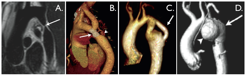
- CT or MRI: these define arch anatomy before surgery and during long-term follow-up (Backer et al., 2023; Isselbacher et al., 2022).
- Four-extremity blood pressures: the ACC/AHA recommends measuring blood pressure in both arms and one leg in patients with CoA (Isselbacher et al., 2022).
Emergency management. For a newborn in shock or with suspected ductal-dependent CoA, standard practice includes:
- Prostaglandin E1 infusion to reopen or maintain the ductus. This can be started on clinical suspicion before echocardiographic confirmation.
- Resuscitation, ventilatory support as needed, and correction of acidosis.
- Urgent transfer to a pediatric cardiac center.
The Pediatrics review notes that when in doubt, assessment by a pediatric cardiologist is mandatory (Hysko et al., 2025). Prostaglandin can cause apnea, so the setting and team for transport matter.
Treatment
- Surgery is the treatment of choice in newborns, infants, and young children. The common technique is resection with an extended end-to-end anastomosis, through a side (thoracotomy) incision or, with arch hypoplasia, through the breastbone. Catheter-based stenting is preferred in older children (over about 25 kg) and adults (Dijkema et al., 2017).
- Some centers use catheter-based stenting as a temporary bridge in critically ill newborns, with later surgery. Balloon angioplasty alone does not complicate later surgical repair, but a stent must be removed during the later operation (Hysko et al., 2025).
- Risk factors for death and recurrent narrowing include poor clinical condition, prostaglandin infusion at the time of surgery, weight under 2.5 kg, small anatomy, associated heart defects, and arch hypoplasia (Hysko et al., 2025).
These risk factors are relevant to causation, because some reflect how sick the child was at diagnosis, while others reflect underlying anatomy present regardless of timing.
Medical Considerations in Evaluating Standards of Care
The legal standard of care is defined by jurisdiction-specific law and expert testimony. Clinical guidelines and literature inform, but do not define, that standard. Expert reviewers commonly examine the following.
Nursery examination.
- Was the femoral pulse exam documented as actually performed?
- Were abnormal or equivocal findings acted on, for example with four-extremity blood pressures or an echocardiogram?
The tertiary-center review concluded that checking peripheral pulses at the first and early follow-up newborn exams, and considering CoA in any patient with hypertension, are needed to improve timely detection (Geggel, 2022).
Pulse oximetry. Was screening done, timed, and interpreted according to the algorithm in effect, with the required follow-up?
Follow-up visits. When a newborn returns with poor feeding, fast breathing, sweating, or poor weight gain, was a cardiovascular cause considered and examined for? Moderate-to-severe heart dysfunction occurred mainly in infants in the first month, who often had fast breathing or poor weight gain (Geggel, 2022).
Telephone triage. Were symptoms such as fast breathing and refusal to feed in a young infant triaged with appropriate urgency?
Emergency care. In a shocked newborn, was ductal-dependent heart disease considered alongside sepsis? Was prostaglandin started without unreasonable delay, and was transfer arranged promptly?
Prenatal follow-through. If CoA was suspected before birth, was the delivery plan followed, including delivery location, serial echocardiograms, and delaying discharge until the ductus closed?
Hypertension in older children and adults. Were arm and leg blood pressures and femoral pulses assessed before the hypertension was attributed to another cause?
Postoperative surveillance. Was recurrent narrowing, hypertension, or aneurysm monitored according to recommended intervals?
Common Allegations in Litigation
Failure to examine femoral pulses
- What plaintiffs often argue
- Template "normal" entries mask an exam that was not done
- What defendants often argue
- Pulses can be normal while the ductus is open
Pulse oximetry error or omission
- What plaintiffs often argue
- Screening omitted, misread, or abnormal results not followed up
- What defendants often argue
- A properly performed screen misses many CoA cases
Missed prenatal diagnosis
- What plaintiffs often argue
- Fetal scan should have raised suspicion
- What defendants often argue
- Prenatal CoA detection is inherently limited
Failure to recognize symptoms
- What plaintiffs often argue
- Poor feeding, sweating, and fast breathing were red flags
- What defendants often argue
- Symptoms are nonspecific and common in healthy newborns
Delayed prostaglandin or transfer
- What plaintiffs often argue
- Shock attributed to sepsis alone
- What defendants often argue
- Presentation mimicked sepsis, and treatment was appropriate once suspected
Misattributed hypertension
- What plaintiffs often argue
- Childhood hypertension called essential or renal without a pulse or blood pressure check
- What defendants often argue
- No signs pointed to CoA at the time
Inadequate postoperative follow-up
- What plaintiffs often argue
- Recurrent narrowing, aneurysm, or hypertension not detected
- What defendants often argue
- Late complications occur despite appropriate surveillance
Documentation deficiencies
- What plaintiffs often argue
- Missing or copied-forward exam findings
- What defendants often argue
- Documentation gaps do not prove the exam was not done
| Allegation | What plaintiffs often argue | What defendants often argue |
|---|---|---|
| Failure to examine femoral pulses | Template "normal" entries mask an exam that was not done | Pulses can be normal while the ductus is open |
| Pulse oximetry error or omission | Screening omitted, misread, or abnormal results not followed up | A properly performed screen misses many CoA cases |
| Missed prenatal diagnosis | Fetal scan should have raised suspicion | Prenatal CoA detection is inherently limited |
| Failure to recognize symptoms | Poor feeding, sweating, and fast breathing were red flags | Symptoms are nonspecific and common in healthy newborns |
| Delayed prostaglandin or transfer | Shock attributed to sepsis alone | Presentation mimicked sepsis, and treatment was appropriate once suspected |
| Misattributed hypertension | Childhood hypertension called essential or renal without a pulse or blood pressure check | No signs pointed to CoA at the time |
| Inadequate postoperative follow-up | Recurrent narrowing, aneurysm, or hypertension not detected | Late complications occur despite appropriate surveillance |
| Documentation deficiencies | Missing or copied-forward exam findings | Documentation gaps do not prove the exam was not done |
Medical Causation
Causation is often the most contested issue. Common considerations include:
- Timing of ductal closure. Deterioration can occur abruptly. Experts often try to determine when the child would most likely have shown detectable findings, and whether earlier diagnosis would most likely have preceded the collapse.
- Shock and organ injury. Kidney injury, necrotizing enterocolitis, liver injury, and brain injury follow low blood flow and acidosis. Their severity often correlates with how long and how deep the shock was.
- Heart function. Severe left ventricular dysfunction is associated with longer ventilation and longer hospital and ICU stays (Hysko et al., 2025). Function often recovers after repair, which affects damages analysis.
- Underlying anatomy. Arch hypoplasia, bicuspid valve, associated defects, low birth weight, and genetic syndromes affect outcomes regardless of diagnostic timing (Hysko et al., 2025).
- Long-term hypertension. Preclinical data suggest that the duration and severity of coarctation predict vascular remodeling, and the conventional 20 mm Hg treatment threshold may be too high to prevent it (Hysko et al., 2025). This is an evolving area. Human evidence linking a delay of days to weeks with lifelong hypertension is limited.
- Probability versus possibility. Courts generally require that causation be established to a "reasonable degree of medical probability," not merely possibility. An expert must explain why a specific injury more likely than not would have been avoided with earlier action.
Long-Term Outcomes
Survival. Survival after surgical repair is excellent.
- In a 341-patient Italian series, 30-day mortality was 1.4%. At 21 years, freedom from death was 93.5% and freedom from aortic reintervention was 93.6% (Padalino et al., "Surgical Repair of Aortic Coarctation in Pediatric Age: A Single Center Two Decades Experience," Journal of Cardiac Surgery, 2019).
- A Dutch series of 292 children reported 96% survival at 15 years (Farag et al., "Aortic Coarctation Repair Through Left Thoracotomy: Results in the Modern Era," European Journal of Cardio-Thoracic Surgery, 2019).
Recurrent narrowing (recoarctation).
- Recoarctation occurred in 9.9% of the Dutch cohort overall, but in 21% after repair in the newborn period, and it occurred as late as 14 years after surgery (Farag et al., 2019).
- The ACC/AHA cites recurrence of about 10% after surgery and about 8% after balloon dilation (Isselbacher et al., 2022).
- Reintervention rates in infants have ranged from 3% to 18.5% (Hysko et al., 2025).
Recoarctation is an expected risk, not proof of surgical error.
Hypertension. Hypertension is the main long-term problem. A 2022 systematic review reported a prevalence of 47.3% (range 20%–70%) after neonatal repair, linked to abnormal vascular remodeling, autonomic dysfunction, residual obstruction, and arch shape (Hysko et al., 2025). Hypertension is especially common during exercise, and ambulatory and exercise testing can help detect it (Isselbacher et al., 2022). Reported rates of antihypertensive treatment vary widely, from 3.0% (Padalino et al., 2019) to 14% (Farag et al., 2019), likely reflecting differences in definitions and follow-up.
Aortic and other complications. Later complications include aneurysm or pseudoaneurysm at the repair site, aortic dissection, and ascending aortic enlargement in patients with bicuspid valves. Adults may have brain aneurysms (Gurvitz et al., 2026; Isselbacher et al., 2022). Despite adequate repair, patients still have reduced life expectancy and more cardiovascular problems later in life (Dijkema et al., 2017).
Lifelong follow-up. Guidelines call for lifelong cardiology follow-up:
- Blood pressures in both arms and one leg (Isselbacher et al., 2022)
- Aortic imaging with MRI or CT, often every 3–5 years once stable (Isselbacher et al., 2022; Mazzolai et al., "2024 ESC Guidelines for the Management of Peripheral Arterial and Aortic Diseases," European Heart Journal, 2024)
- Consideration of screening for brain aneurysms in adults (Isselbacher et al., 2022)
Neurodevelopment. Children with isolated CoA repaired without shock generally have favorable outcomes. Those who experienced profound shock, cardiac arrest, or brain injury may have lasting neurodevelopmental effects. CoA-specific data are limited, so causation opinions should rely on the child's individual imaging and testing.
Medical Records That Often Matter
- Prenatal records: ultrasound reports and images, fetal echocardiograms, referral letters, and delivery plans
- Nursery records: physical exam (especially femoral pulses), vital signs, blood pressures, and pulse oximetry logs with times and extremities tested
- Nursing notes: feeding, color, breathing rate, and parental concerns
- Outpatient and telephone records: weight checks, triage call logs, after-hours notes, and recordings where available
- Emergency department and transport records: timeline of vital signs, lactate and blood gases, prostaglandin start time, and medications
- Echocardiograms: both the reports and the original images, ideally reviewed by an independent pediatric cardiologist
- Operative, ICU, and catheterization records
- Follow-up records: blood pressures, imaging, and neurodevelopmental and kidney evaluations
Electronic health record audit trails can show when exam templates were completed and whether entries were copied forward.
Common Misconceptions
- "A passed pulse oximetry screen rules out serious heart disease." Sensitivity for coarctation may be as low as 21% (Oster et al., 2025).
- "A normal prenatal scan rules out CoA." Prenatal detection of CoA remains difficult (Dijkema et al., 2017).
- "Normal femoral pulses at birth exclude CoA." An open ductus can mask the obstruction until it closes (Hysko et al., 2025).
- "No murmur means no heart disease." Many critical cases have no significant murmur.
- "An early normal echocardiogram excludes CoA." Ductal flow can mask severe narrowing (Hysko et al., 2025).
- "Successful repair means a cure." Hypertension, recoarctation, and aortic complications require lifelong surveillance (Isselbacher et al., 2022).
- "Delayed diagnosis always causes the injury." Outcomes depend heavily on anatomy, associated defects, and how quickly the decompensation evolved.
The Value of Pediatric Cardiology Expertise
CoA cases involve the following areas of expertise:
- Newborn circulatory transition and the behavior of the ductus
- The limits of fetal echocardiography and pulse oximetry screening
- Independent interpretation of echocardiographic and CT images
- Surgical and catheter treatment options
- Long-term outcome literature
Opinions on general pediatric, nursery, or emergency care may require experts in those fields. Causation opinions often require pediatric cardiology input. Separating complications caused by delay from those inherent to the anatomy is a specialized task.
Key Takeaways for Attorneys
- Coarctation is among the heart defects most often diagnosed after nursery discharge. Nearly half of isolated cases went home undiagnosed in one population study.
- Closure of the ductus arteriosus can turn a well-appearing newborn into a baby in shock within days to weeks.
- Pulse oximetry detects CoA poorly, with sensitivity as low as 21%. A passed screen is not exculpatory or inculpatory on its own.
- Prenatal detection of CoA is limited. Follow-through on prenatal suspicion is often more relevant than the scan itself.
- The femoral pulse exam and four-extremity blood pressures are central. Their documentation, and its authenticity, are frequently examined.
- Nonspecific newborn symptoms (poor feeding, sweating, fast breathing) are a common point of dispute about when CoA should have been suspected.
- In a newborn in shock, consideration of ductal-dependent heart disease and timely prostaglandin are key review points.
- Hypertension in children should prompt consideration of CoA. Misattribution to other causes is a recognized pattern of delay.
- Recoarctation and hypertension occur even after excellent repair and do not by themselves indicate substandard care.
- Causation requires distinguishing injury from delay versus injury from anatomy and natural disease course, to a reasonable degree of medical probability.
This article is for educational purposes only and does not constitute legal advice or a medical opinion about any individual patient or case.
References
- Lannering et al. Late Diagnosis of Coarctation Despite Prenatal Ultrasound and Postnatal Pulse Oximetry. Pediatrics. 2015.
- Isselbacher et al. 2022 ACC/AHA Guideline for the Diagnosis and Management of Aortic Disease. Circulation/JACC. 2022.
- Gurvitz et al. 2025 ACC/AHA/HRS/ISACHD/SCAI Guideline for the Management of Adults With Congenital Heart Disease. Journal of the American College of Cardiology. 2026.
- Dijkema et al. Diagnosis, Imaging and Clinical Management of Aortic Coarctation. Heart. 2017.
- Hysko et al. Advances in the Treatment of Neonatal Coarctation of the Aorta. Pediatrics. 2025.
- Backer et al. Coarctation of the Aorta. Pediatric Cardiac Surgery, 5th ed. 2023.
- Oster et al. Newborn Screening for Critical Congenital Heart Disease: A New Algorithm and Other Updated Recommendations. Pediatrics. 2025.
- Geggel. Coarctation of the Aorta: Delay in Diagnosis and Referral Basis From Infancy to Adulthood. Journal of Pediatrics. 2022.
- Jullien. Newborn Pulse Oximetry Screening for Critical Congenital Heart Defects. BMC Pediatrics. 2021.
- Martin et al. Updated Strategies for Pulse Oximetry Screening for Critical Congenital Heart Disease. Pediatrics. 2020.
- Padalino et al. Surgical Repair of Aortic Coarctation in Pediatric Age: A Single Center Two Decades Experience. Journal of Cardiac Surgery. 2019.
- Farag et al. Aortic Coarctation Repair Through Left Thoracotomy: Results in the Modern Era. European Journal of Cardio-Thoracic Surgery. 2019.
- Mazzolai et al. 2024 ESC Guidelines for the Management of Peripheral Arterial and Aortic Diseases. European Heart Journal. 2024.
Educational Disclaimer
This article is provided for educational purposes only and does not constitute legal or medical advice. Every case is unique and should be evaluated on its individual facts and medical records. Reading this article does not create an expert-client relationship.
About the Author
Asif Masood, MD, MSc is double board-certified in General Pediatrics and Pediatric Cardiology. Expert witness services in general pediatrics and pediatric & congenital cardiology, complemented by independent, non-testimony medical consulting across a broad range of medical specialties. Through The Verdict MD, he serves attorneys, law firms, insurers, and healthcare organizations nationwide.
Learn about Dr. Masood's pediatric cardiology expert witness services for independent review of the medical questions in your case.