Pediatric & Congenital Cardiology
Double Outlet Right Ventricle (DORV): A Medical-Legal Guide for Attorneys
- Published
- November 10, 2026
- Last Reviewed
- November 10, 2026
- Author
- Asif Masood, MD, MSc
- Reading Time
- 16 min
- Category
- Pediatric & Congenital Cardiology
Double outlet right ventricle (DORV) is not one disease. It is a family of heart defects that share one feature: both great arteries (the aorta and the pulmonary artery) arise wholly or mostly from the right ventricle. The only outlet for the left ventricle is a hole between the ventricles, a ventricular septal defect (VSD) (Gurvitz et al., Journal of the American College of Cardiology, 2026). This article explains the anatomy, diagnosis, treatment, and outcomes of DORV in plain language, and describes the medical issues most often examined in litigation. It is educational. It does not offer legal advice or opinions on liability in any specific case, and it does not suggest that any particular finding automatically means negligence.
Why the Subtype Matters
DORV occurs in about 0.1–0.2 per 1,000 live births (Young et al., Prenatal Diagnosis, 2021). Depending on its anatomy, a child with DORV may behave like a child with a large VSD, with tetralogy of Fallot, with transposition of the great arteries, or with a single functioning ventricle (Young et al., 2021). This range matters in litigation. The expected symptoms, the urgency of diagnosis, the type of surgery, and the expected outcome all depend on which form of DORV the child has. A case analysis that treats "DORV" as a single condition with a single prognosis will usually be wrong.
Hypothetical Clinical Scenario
The following case is fictional and is presented only for educational purposes.
At 20 weeks of pregnancy, a 31-year-old woman has a routine anatomy ultrasound at a community imaging center. The report describes a "normal four-chamber heart." The outflow tract views are listed as "suboptimal due to fetal position." No follow-up cardiac imaging or fetal echocardiogram is recommended.
The baby boy is born at term at a community hospital. His oxygen saturation on the newborn pulse oximetry screen at 26 hours is 93% in the foot and 95% in the right hand. The screen is repeated an hour later with the same results. A third reading, documented as 95% in both, is recorded as a "pass." He is discharged on day 2.
At 2 weeks, his pediatrician notes a loud murmur and documents "mild duskiness with crying." A cardiology referral is placed as "routine," with an appointment 5 weeks away. At 5 weeks, he presents to an emergency department with poor feeding, fast breathing, and an oxygen saturation of 78%. An echocardiogram shows DORV with a VSD beneath the pulmonary artery (Taussig-Bing type), with transposition-like physiology and coarctation of the aorta. He is transferred to a children's heart center.
Because he is now older than the usual window for an arterial switch, the surgical team must assess whether his left ventricle is still strong enough to support the body. He undergoes an arterial switch with VSD closure and arch repair. His postoperative course is prolonged, and he later needs a reoperation for narrowing below the new aortic valve.
The scenario raises several questions that experts would examine separately: whether the prenatal outflow views should have prompted further imaging; how the pulse oximetry screen was interpreted; whether the referral urgency fit the findings at 2 weeks; and whether, and how much, the timing of diagnosis changed the surgical result. None of these questions answers itself, and each requires separate medical analysis.
Understanding the Anatomy
In a normal heart, the left ventricle pumps oxygen-rich blood to the aorta and the body, and the right ventricle pumps oxygen-poor blood to the pulmonary artery and the lungs. In DORV, both arteries come off the right ventricle. Blood from the left ventricle can only leave through the VSD.
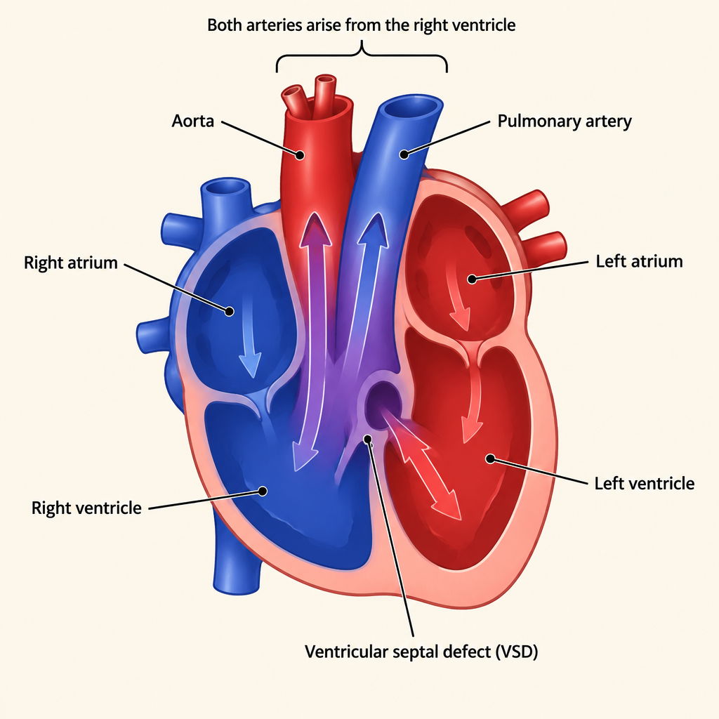
Where the VSD sits relative to the two great arteries largely decides how the child behaves and what surgery is needed. Cardiologists usually describe four VSD locations (Lopez et al., Echocardiography in Pediatric and Congenital Heart Disease, 2021; Mavroudis et al., Pediatric Cardiac Surgery, 2023):
- Subaortic VSD (the most common). The hole sits under the aorta. Without pulmonary stenosis, the child behaves like a child with a large VSD: excess lung blood flow and heart failure over the first weeks to months. With pulmonary stenosis, the child behaves like one with tetralogy of Fallot and may be blue (cyanotic).
- Subpulmonary VSD (Taussig-Bing anomaly). The hole sits under the pulmonary artery. Oxygen-rich blood goes mostly to the lungs and oxygen-poor blood mostly to the body, as in transposition of the great arteries. These infants are often cyanotic from early life and often have narrowing of the aortic arch (coarctation) (Urbina et al., NeoReviews, 2025).
- Doubly committed VSD. The hole sits under both arteries.
- Noncommitted (remote) VSD. The hole is far from both arteries. This is the hardest form to repair with two ventricles, because the surgeon must build a long internal tunnel to connect the left ventricle to an artery (Lopez et al., 2021).
"Complex" DORV. Additional defects make repair harder and outcomes less predictable. The 2025 ACC/AHA adult congenital heart disease guideline lists atrial isomerism (heterotaxy), atrioventricular septal defect, multiple VSDs, an underdeveloped right or left ventricle, straddling valves, and major pulmonary artery abnormalities. These features influence whether a two-ventricle repair is possible at all (Gurvitz et al., 2026).
Associated conditions. DORV diagnosed before birth frequently comes with other problems. In one fetal series, 91% had major additional heart defects, 67% had defects outside the heart, and 29% had chromosomal or syndromic conditions (Gottschalk et al., Journal of Perinatal Medicine, 2019). These associated conditions often drive outcome independently of any delay in diagnosis, and they are central to causation analysis.
Prenatal Diagnosis
Why DORV can be missed. The standard four-chamber view of the fetal heart is often normal in DORV, because the abnormality is in how the arteries leave the heart. Recognizing DORV requires the outflow tract views. A Pediatrics study of US prenatal detection found low detection rates for transposition, tetralogy, and DORV, attributed partly to reliance on the four-chamber view alone. Obstetric guidelines have since been updated to include outflow tract views in the second-trimester anatomy scan (Quartermain et al., Pediatrics, 2015).
Detection rates. Prenatal detection has improved. Population studies in the current era report that 60–78% of DORV cases are now detected before birth (Young et al., 2021). That still leaves a meaningful share that is not detected even in good systems. Detection varies with sonographer training, fetal position, maternal body habitus, gestational age, and equipment.
Accuracy once suspected. When a fetus is referred for a fetal echocardiogram by an experienced team, diagnostic accuracy is high: 96.3% for the diagnosis of DORV in one series, and about 97% in another (Gottschalk et al., 2019; Öcal et al., Echocardiography, 2024). Prediction of the exact subtype and of the eventual surgical plan is less reliable for the most complex forms.
Medicolegal relevance. Prenatal cases usually turn on questions such as:
- Were outflow tract views obtained, and were they adequate?
- If views were "suboptimal" or "limited," was repeat imaging or a fetal echocardiogram recommended?
- Were risk factors present that warranted fetal echocardiography (for example, maternal diabetes, family history, other fetal anomalies, or abnormal genetic screening)?
- Was an abnormal finding communicated to the ordering obstetrician and the family?
Prenatal diagnosis allows delivery planning, genetic counseling, and in some cases a decision about whether to continue the pregnancy. In "wrongful birth" claims, which exist only in some jurisdictions, the associated extracardiac and genetic conditions are often as important as the heart defect itself. Not every missed prenatal diagnosis reflects a deviation from accepted practice; some defects cannot be seen even with good technique.
Postnatal Presentation and Recognition
The newborn's course depends on the subtype:
- Transposition-type (Taussig-Bing). Cyanosis may be obvious within hours to days. When an aortic arch obstruction is also present, the baby may depend on the ductus arteriosus and can collapse when it closes, often in the first 1–2 weeks.
- Tetralogy-type. Cyanosis ranges from mild to severe depending on the degree of pulmonary stenosis. "Spells" with sudden worsening cyanosis can occur.
- VSD-type (no pulmonary stenosis). The baby may look well at birth. As lung resistance falls over the first weeks, heart failure develops: fast breathing, sweating with feeds, poor weight gain, and a murmur. Untreated, persistent excess lung blood flow can damage the lung blood vessels over time.
Pulse oximetry screening. Newborn pulse oximetry screening is designed to detect critical heart disease that causes low oxygen levels. It catches many transposition-type and tetralogy-type cases. It may miss the VSD-type form, where oxygen levels are often normal. Screening results in the borderline range (in the scenario above, 93–95%) require correct application of the screening algorithm; misinterpreting a borderline result is a recurring issue in congenital heart disease litigation.
Murmurs. Many forms of DORV produce a murmur, but its loudness does not track severity. A murmur in a newborn may be called "innocent" early; persistence, change in character, or accompanying symptoms usually prompt further evaluation.
Diagnostic Evaluation
Echocardiography is the primary diagnostic test. It identifies both great arteries arising from the right ventricle, the VSD's position, any outflow obstruction, arch abnormalities, and valve problems. For complex forms, surgical planning often adds cardiac CT or MRI, and sometimes three-dimensional modeling, to decide whether the VSD can be connected to an artery through a tunnel (Urbina et al., 2025).
When a newborn has cyanosis or suspected ductal dependence, the accepted response includes starting prostaglandin E1 to keep the ductus open, and transferring the baby to a center that offers pediatric cardiology and congenital heart surgery. Timeliness of these steps is often examined in litigation.
Treatment
Individualized repair. Because the anatomy varies so widely, surgical approach and timing must be individualized (Urbina et al., 2025). Most children can achieve a biventricular (two-ventricle) repair, but some need staged procedures or single-ventricle palliation.
Main surgical strategies (Gurvitz et al., 2026; Urbina et al., 2025):
- Intraventricular tunnel (baffle) repair. For subaortic or doubly committed VSDs, a patch tunnel routes left-ventricle blood through the VSD to the aorta. Pulmonary stenosis is relieved if present.
- Arterial switch with VSD tunneling. For the Taussig-Bing form, the VSD is tunneled to the pulmonary valve and the arteries are switched, as in transposition. These repairs are often done in the newborn period, before the left ventricle loses strength, especially when an aortic arch obstruction is present (Urbina et al., 2025).
- Rastelli, REV, or root translocation procedures. For subpulmonary VSD with pulmonary stenosis, or other complex forms. The Rastelli procedure uses a conduit from the right ventricle to the pulmonary artery, and conduits need replacement as children grow.
- Single-ventricle (Fontan) pathway. When a two-ventricle repair is not feasible, for example with a severely underdeveloped ventricle or straddling valves.
Temporizing procedures. Some infants first need a pulmonary artery band to limit excess lung blood flow, or a shunt or stent to add lung blood flow. Prematurity, growth restriction, genetic syndromes, multiple heart defects, or major non-cardiac problems often favor palliation before complete repair (Urbina et al., 2025).
Timing. For the tetralogy-type, repair is generally advised by about 6 months of age when an intraventricular repair is clearly feasible. Some centers favor early primary repair for nearly all patients, while others use an initial shunt in selected children (Mavroudis et al., 2023). This is an area where reasonable, well-trained surgeons differ. Center-specific practices within the accepted range should not be confused with deviations from accepted care.
Outcomes
Survival. Contemporary results are generally good but vary by subtype and complexity:
- In 238 patients with biventricular repair at a US center, 5-year survival was 89.0% and freedom from reoperation for left ventricular outflow obstruction was 84% (Oladunjoye et al., Journal of Thoracic and Cardiovascular Surgery, 2020).
- In a national Norwegian cohort of 93 children, survival was 89.5% at 10 years, and freedom from surgical reintervention was 69.4% at 10 years. Associated atrioventricular septal defect substantially increased mortality risk (Holten-Andersen et al., European Journal of Cardio-Thoracic Surgery, 2022).
- In 1,135 patients at a large Chinese center, 15-year survival was 92.7%. Transposition-type anatomy and palliative procedures were independently associated with higher mortality (Pang et al., European Journal of Cardio-Thoracic Surgery, 2025).
- In 380 biventricular repairs, preoperative pulmonary hypertension was the only risk factor for early death (Li et al., Journal of Thoracic and Cardiovascular Surgery, 2014).
Reintervention is expected, not exceptional. Many children with DORV will need more surgery or catheter procedures over their lifetime. Common reasons include:
- Narrowing of the tunnel or below the aortic valve (left ventricular outflow tract obstruction). This is the leading cause of reoperation after biventricular repair and can develop late (Li et al., 2014; Gurvitz et al., 2026).
- Conduit replacement after Rastelli-type repairs.
- Residual VSD patch leaks, which the adult guideline says should be looked for routinely (Gurvitz et al., 2026).
- Problems specific to the arterial switch (coronary or neo-aortic issues) in transposition-type cases.
- Heart block and arrhythmias, related to the location of the conduction system near the VSD.
A later reoperation is therefore not, by itself, evidence of a surgical error.
Lifelong care. Adults with repaired DORV are managed according to the repair they received, for example like tetralogy patients with a conduit or transposition patients after arterial switch (Gurvitz et al., 2026). They need lifelong follow-up with a congenital heart specialist.
Neurodevelopment. Children with complex congenital heart disease, especially those with cyanosis, genetic syndromes, or multiple surgeries, are at increased risk of developmental, learning, and attention problems. Separating injury caused by an alleged delay from these baseline risks is one of the hardest parts of causation analysis.
Targets for Litigation: Issues Commonly Examined
The following are areas that commonly draw scrutiny in DORV cases. Each requires a fact-specific medical review; none automatically establishes a deviation from accepted practice or causation.
1. Prenatal imaging
- Missing or inadequate outflow tract views on the anatomy scan.
- "Limited" or "suboptimal" views without a recommendation for repeat imaging or fetal echocardiography.
- Failure to refer for fetal echocardiography when risk factors were present.
- Failure to communicate abnormal findings to the obstetrician or family.
- Inadequate genetic counseling or testing after a prenatal diagnosis.
2. Newborn nursery care
- Pulse oximetry screening not performed, performed incorrectly, or misread (especially borderline or repeat results).
- Cyanosis, fast breathing, or poor feeding not recognized or not acted on before discharge.
- A murmur noted but not followed up or communicated to the outpatient pediatrician.
3. Outpatient and emergency care
- Symptoms of heart failure (fast breathing, sweating with feeds, poor weight gain) attributed to feeding problems, reflux, or a respiratory infection.
- Persistent or new murmurs labeled "innocent" without reassessment.
- Referral urgency that does not fit the findings, such as a "routine" referral for an infant with cyanosis.
- Delayed prostaglandin, delayed transfer, or delayed cardiology contact for a cyanotic or shocked infant.
4. Cardiology diagnosis
- Incomplete or misread echocardiograms, including misclassifying the subtype, missing arch obstruction, coronary anomalies, straddling valves, or additional VSDs.
- Failure to obtain advanced imaging (CT, MRI) when the anatomy was unclear and surgical planning depended on it.
5. Surgical decision-making and timing
- Choice between biventricular repair, staged repair, and single-ventricle palliation.
- Timing of repair, particularly for transposition-type DORV, where delay can weaken the left ventricle, and for VSD-type DORV, where delay risks pulmonary vascular disease.
- Adequacy of informed consent, including discussion of alternatives, expected reoperations, and center-specific results.
- Whether a case of this complexity was referred to a center with appropriate experience.
6. Surgical technique and complications
- Tunnel or outflow obstruction, residual VSD, heart block requiring a pacemaker, and coronary complications after an arterial switch.
- Many of these are known risks of correctly performed surgery. Analysis focuses on whether a complication was recognized and managed appropriately, not just whether it occurred.
7. Postoperative and long-term follow-up
- Delayed recognition of low cardiac output, arrhythmias, or residual lesions in the ICU.
- Missed surveillance for progressive outflow obstruction, conduit failure, or arrhythmia.
- Gaps in transition from pediatric to adult congenital heart care.
Medical Causation
Causation in DORV cases is often more contested than the question of whether care fell short. Factors experts commonly weigh include:
- Subtype and complexity. A remote VSD, heterotaxy, an underdeveloped ventricle, or an atrioventricular septal defect may limit outcome regardless of when diagnosis occurred.
- Associated genetic and extracardiac conditions. These independently affect survival and neurodevelopment.
- Physiologic consequences of delay. Examples include severe hypoxemia, acidosis, shock from ductal closure, left ventricular deconditioning in transposition-type DORV, and pulmonary vascular disease in unrepaired VSD-type DORV. Documented end-organ injury (for example, brain, kidney, or bowel injury at the time of collapse) is often the strongest link between a delay and harm.
- Whether earlier diagnosis would have changed management. Prenatal diagnosis may have changed delivery location and early stabilization, but the eventual operation may have been identical.
- Expected reintervention. Because many children need more surgery regardless of care quality, attributing later procedures to an alleged deviation requires specific medical reasoning.
- Probability versus possibility. Causation opinions must generally be stated to a reasonable degree of medical probability (more likely than not), not as a mere possibility. The legal standard is jurisdiction-specific.
Medical Records That Often Matter
- Prenatal ultrasound images (not only reports), fetal echocardiograms, and genetic testing.
- Delivery records and newborn nursery notes, including vital signs and nursing assessments.
- Pulse oximetry screening logs with every reading, the site, the time, and the interpretation.
- Pediatric office notes, growth charts, and referral documents showing the urgency assigned.
- Emergency department, transport, and prostaglandin administration records.
- All echocardiograms, CT, MRI, and catheterization studies, with original images when possible.
- Surgical conference notes, consent forms, operative reports, and perfusion records.
- ICU records, discharge summaries, and long-term cardiology and neurodevelopmental follow-up.
Common Misconceptions
- "A normal four-chamber view rules out serious heart disease." DORV often has a normal four-chamber view; the outflow views are needed (Quartermain et al., 2015).
- "Passing pulse oximetry rules out DORV." Forms with normal oxygen levels, such as VSD-type DORV, can pass.
- "DORV has a single prognosis." Outcomes vary widely by subtype, associated anomalies, and repair type (Pang et al., 2025; Holten-Andersen et al., 2022).
- "A reoperation means the first surgery was done wrong." Reintervention is common and expected after many DORV repairs (Holten-Andersen et al., 2022; Li et al., 2014).
- "Single-ventricle palliation means the surgeon chose wrongly." For some anatomy, a two-ventricle repair is not feasible or carries more risk.
The Value of Specialized Expertise
DORV cases cross several specialties: maternal-fetal medicine and fetal cardiology for prenatal issues, neonatology and general pediatrics for newborn and outpatient recognition, pediatric cardiology and cardiac imaging for diagnosis, and congenital heart surgery for repair decisions. Accurate review requires matching the expert to the issue. A reviewer must understand the specific subtype, the published outcomes for that subtype, and the range of accepted surgical approaches, so that preventable harm can be separated from the inherent risks of a complex malformation.
Key Takeaways for Attorneys
- DORV is a spectrum of defects, not one condition. The VSD's location largely determines symptoms, surgery, and prognosis.
- Prenatal detection now reaches about 60–78% in population studies, but the four-chamber view is often normal; the outflow tract views are key.
- Transposition-type and tetralogy-type DORV usually cause cyanosis; VSD-type DORV may present weeks later with heart failure and can pass pulse oximetry screening.
- Prostaglandin and urgent transfer are central for cyanotic or ductal-dependent newborns.
- Surgical strategy and timing are individualized, and accepted practice includes more than one reasonable approach.
- Contemporary long-term survival is roughly 86–93% in large series, lower with complex anatomy, atrioventricular septal defect, or palliation.
- Reintervention, especially for outflow tract obstruction or conduit failure, is common and does not by itself indicate error.
- Associated genetic and extracardiac conditions frequently drive outcome and must be considered in causation.
- The strongest causation cases usually document a physiologic injury, such as shock, acidosis, or end-organ damage, linked in time to a delay.
- Standard-of-care conclusions are legal determinations. Medical experts inform them by comparing the care given with evidence-based practice at the time.
References
- Gurvitz et al., Journal of the American College of Cardiology, 2026.
- Young et al., Prenatal Diagnosis, 2021.
- Lopez et al., Echocardiography in Pediatric and Congenital Heart Disease, 2021.
- Mavroudis et al., Pediatric Cardiac Surgery, 2023.
- Urbina et al., NeoReviews, 2025.
- Gottschalk et al., Journal of Perinatal Medicine, 2019.
- Quartermain et al., Pediatrics, 2015.
- Öcal et al., Echocardiography, 2024.
- Oladunjoye et al., Journal of Thoracic and Cardiovascular Surgery, 2020.
- Holten-Andersen et al., European Journal of Cardio-Thoracic Surgery, 2022.
- Pang et al., European Journal of Cardio-Thoracic Surgery, 2025.
- Li et al., Journal of Thoracic and Cardiovascular Surgery, 2014.
Educational Disclaimer
This article is provided for educational purposes only and does not constitute legal or medical advice. Every case is unique and should be evaluated on its individual facts and medical records. Reading this article does not create an expert-client relationship.
About the Author
Asif Masood, MD, MSc is double board-certified in General Pediatrics and Pediatric Cardiology. Expert witness services in general pediatrics and pediatric & congenital cardiology, complemented by independent, non-testimony medical consulting across a broad range of medical specialties. Through The Verdict MD, he serves attorneys, law firms, insurers, and healthcare organizations nationwide.
Learn about Dr. Masood's pediatric cardiology expert witness services for independent review of the medical questions in your case.