Pediatric & Congenital Cardiology
Fetal Echocardiography: A Medical-Legal Guide for Attorneys
- Published
- October 31, 2026
- Last Reviewed
- October 31, 2026
- Author
- Asif Masood, MD, MSc
- Reading Time
- 8 min
- Category
- Pediatric & Congenital Cardiology
Congenital heart disease (CHD) is the most common serious birth defect. Prenatal diagnosis gives families time to consider their options, get genetic testing, and plan delivery at a center that can care for the newborn's heart right away. When a baby is born with an undiagnosed heart defect, families often ask: "Why wasn't this seen on the ultrasound?" That question leads to many claims of wrongful birth, missed diagnosis, and delivery-planning failures. To evaluate these cases, attorneys need to understand one key distinction: a routine obstetric screening ultrasound is a different test from a fetal echocardiogram, and each has known limits.
Clinical Vignette (Hypothetical)
A 31-year-old woman with type 1 diabetes before pregnancy has a routine anatomy ultrasound at 20 weeks. The report says "four-chamber heart view normal." It does not mention the outflow tracts (the great arteries leaving the heart), and no follow-up imaging is ordered. Nobody refers her for a fetal echocardiogram.
The baby is born at a community hospital without pediatric cardiology. At 18 hours of age the baby becomes blue (cyanotic) and does not improve with oxygen. An echocardiogram shows transposition of the great arteries with limited mixing between the two circulations. The baby needs emergency transfer and has a period of severe low oxygen before stabilizing.
Potential plaintiff issues
- Pregestational diabetes is a recognized indication for fetal echocardiography.
- The screening ultrasound did not document the outflow-tract views.
- Delivery happened at a hospital not equipped for the baby's needs.
Potential defense issues
- Transposition is one of the lesions most often missed before birth, even by careful examiners.
- The four-chamber view usually looks normal in transposition.
- Even with a prenatal diagnosis, the baby might still have needed an emergency procedure, so the effect of the delay on the outcome has to be proven.
Screening Ultrasound vs. Fetal Echocardiogram
Purpose
- Routine Obstetric Anatomy Ultrasound
- Screening of all pregnancies
- Fetal Echocardiogram
- Detailed diagnostic study of the heart
Who performs it
- Routine Obstetric Anatomy Ultrasound
- Sonographers, obstetricians, maternal-fetal medicine
- Fetal Echocardiogram
- Usually pediatric/fetal cardiologists
Typical timing
- Routine Obstetric Anatomy Ultrasound
- About 18–22 weeks
- Fetal Echocardiogram
- About 18–22 weeks; earlier (12–16 weeks) in high-risk cases
Cardiac views
- Routine Obstetric Anatomy Ultrasound
- Four-chamber, outflow tracts, three-vessel views (when feasible)
- Fetal Echocardiogram
- Full anatomy, blood flow, and rhythm
Detection of major CHD
- Routine Obstetric Anatomy Ultrasound
- Highly variable
- Fetal Echocardiogram
- More than 90% of complex CHD in experienced hands
| Feature | Routine Obstetric Anatomy Ultrasound | Fetal Echocardiogram |
|---|---|---|
| Purpose | Screening of all pregnancies | Detailed diagnostic study of the heart |
| Who performs it | Sonographers, obstetricians, maternal-fetal medicine | Usually pediatric/fetal cardiologists |
| Typical timing | About 18–22 weeks | About 18–22 weeks; earlier (12–16 weeks) in high-risk cases |
| Cardiac views | Four-chamber, outflow tracts, three-vessel views (when feasible) | Full anatomy, blood flow, and rhythm |
| Detection of major CHD | Highly variable | More than 90% of complex CHD in experienced hands |
US practice guidelines (AIUM/ACR/ACOG/SMFM) say the routine second-trimester scan should include the four-chamber and outflow-tract views, and the three-vessel views if technically possible.
The Standard Cardiac Screening Views
The standard screening exam moves upward through the baby's chest in five views:
- Abdominal (situs) view: Shows the stomach and major vessels in their normal positions.
- Four-chamber view: Shows that the four heart chambers are balanced. It detects only about half of major defects.
- Left ventricular outflow tract: Shows the aorta leaving the left ventricle.
- Pulmonary artery / three-vessel view: Shows the pulmonary artery and how it relates to the aorta and the superior vena cava.
- Three-vessel and trachea view: Shows the aortic arch and the ductus arteriosus together.
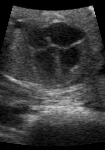
Defects of the outflow tracts and great arteries (for example, transposition and tetralogy of Fallot) can look normal on the four-chamber view. Whether views 3–5 were obtained and documented is often the central record question.
Who Should Be Referred for Fetal Echocardiography?
Guidelines from the American Heart Association (AHA), the American Society of Echocardiography (ASE), and the International Society of Ultrasound in Obstetrics and Gynecology (ISUOG) generally list these indications:
Strongest and most widely accepted
- An abnormal or incomplete heart view on screening ultrasound. Every fetus with an abnormal cardiac screen should have a detailed fetal echocardiogram. Referral is also appropriate when normal heart structures cannot be confirmed.
- A parent or sibling with CHD. AHA rates this Class I: it should be performed.
- Diabetes present before pregnancy, or diabetes diagnosed in the first trimester.
- Increased nuchal translucency (≥3.5 mm), a fluid measurement at the back of the fetal neck.
- Monochorionic twins (twins sharing one placenta).
- Abnormal fetal chromosomes, fluid buildup in the fetus (hydrops), or other major birth defects.
- Mother positive for anti-Ro/SSA or anti-La/SSB antibodies (lupus or Sjögren's), which calls for serial monitoring for heart block.
- First-trimester rubella, poorly controlled phenylketonuria, or specific medication exposures.
Areas where guidelines disagree
- A second-degree relative with CHD (AHA: "may be considered").
- Gestational diabetes. AHA suggests considering a third-trimester study if HbA1c is above 6%.
Guideline strength matters in litigation. A "Class I" indication carries far more weight than a "may be considered" recommendation.
How Accurate Is Prenatal Detection?
- Only about 10% of babies with CHD have a known risk factor. Most defects occur in low-risk pregnancies, so the screening ultrasound is the main safety net.
- In US studies, almost all mothers of babies with serious CHD had a second- or third-trimester ultrasound, yet only about 30% of the defects were diagnosed before birth.
- A four-chamber view alone detects about 50% of major defects. Adding the outflow-tract and three-vessel views can raise detection to as high as 90%.
- Detection varies by defect. Single-ventricle defects are found most often. Transposition and coarctation of the aorta have the lowest prenatal detection rates. In one Swedish population study, detection ranged from 9% to 82% depending on the defect.
- Fetal echocardiography in experienced hands identifies more than 90% of complex CHD. It may still miss small ASDs or VSDs and may underestimate how small the aortic arch or a ventricle is.
Defects That Can Change or Appear Later in Pregnancy
A normal mid-pregnancy exam does not guarantee a normal heart at birth. Some conditions develop or worsen over time:
- Narrowing of a heart valve that leads to an underdeveloped left heart
- Coarctation of the aorta, which is often only suspected and confirmed after birth
- Cardiomyopathy and thickening of the heart muscle (for example, with maternal diabetes)
- Heart rhythm problems, which often do not appear until after 25–26 weeks
The AHA expressly acknowledges that screening at 18–22 weeks may miss disease that progresses later in pregnancy.
Common Targets for Litigation
- Failure to obtain or document the recommended cardiac views on the anatomy scan.
- Failure to refer when a guideline indication was present, especially pregestational diabetes, a family history of CHD, increased nuchal translucency, or monochorionic twins.
- Failure to act on an abnormal or incomplete scan (for example, "heart views suboptimal" with no repeat imaging).
- Misinterpretation of images that, on review, show the abnormality.
- Delayed referral. The AHA recommends evaluation as soon as feasible, and same-day or next-day referral for defects that threaten the fetus's circulation.
- Inadequate follow-up of a known defect. Lesions that can progress need serial studies, including at least one late-pregnancy study for defects that will need early intervention.
- Failure to plan delivery at an appropriate center for defects that depend on the ductus arteriosus or limited mixing, such as transposition or HLHS with a restrictive atrial septum.
- Counseling failures. These include not offering genetic testing or not explaining options in time for decisions about the pregnancy (wrongful birth claims, which vary by state).
- Communication breakdowns between the sonographer, the radiologist or maternal-fetal medicine specialist, the obstetrician, and the cardiologist.
Common Defense Themes
- Screening is not diagnosis. Even well-performed screening misses a substantial share of defects.
- Defect-specific detectability. Transposition, coarctation, total anomalous pulmonary venous return, and small septal defects are known to be hard to see before birth.
- Technical limits. These include maternal body habitus, fetal position, gestational age, and amniotic fluid volume.
- No indication was present. Most affected pregnancies have no risk factor that would trigger referral.
- Progressive disease. The heart may truly have looked normal at the time of the scan.
- Lack of causation. Even with a prenatal diagnosis, the defect, the need for surgery, and many outcomes would have been the same.
- Guideline strength. A "may be considered" recommendation is not a mandatory standard of care.
Causation: The Central Question
Showing that a defect was missed is not enough. The questions that decide these cases include:
- Was the defect detectable at that gestational age with that exam?
- What would a timely diagnosis have changed? The choice to continue the pregnancy (wrongful birth, where recognized); delivery location and immediate prostaglandin or a catheter procedure (balloon atrial septostomy); and avoiding collapse, acidosis, or prolonged low oxygen before diagnosis.
- Did the time without a diagnosis cause injury (brain injury, organ injury, death), or did the defect itself cause the outcome?
Causation is often strongest when the defect depends on the ductus arteriosus or on limited mixing, and the baby collapsed after an unplanned delivery. It is often weakest when the outcome would have been the same with a prenatal diagnosis.
Medical Records That Matter
- Full obstetric ultrasound reports and the stored images and video clips, not just the written report
- Documentation of which cardiac views were obtained, and any "suboptimal" or "limited" notes
- Maternal history: diabetes, family history, medications, autoimmune antibodies
- Nuchal translucency and genetic screening results
- Referral orders, scheduling records, and fetal echocardiogram reports
- Counseling notes and genetic testing offers
- Delivery planning documents
- Newborn records: pulse oximetry screening, timing of cyanosis, prostaglandin start time, transfer timeline
Stored image review is often decisive. It can show either that the abnormality was visible or that the required views were never obtained.
Common Misconceptions
- "A normal anatomy scan means the heart is normal." False. Screening misses many defects.
- "Every heart defect is visible before birth." False. Some defects are inherently hard to detect or develop later.
- "Any missed diagnosis is malpractice." False. The standard is reasonable care, not perfect detection.
- "A prenatal diagnosis would have prevented the defect's consequences." Often false. It mainly changes counseling, delivery planning, and the immediate newborn course.
The Value of Specialty Expertise
These cases cross obstetrics, maternal-fetal medicine, radiology, and pediatric cardiology. A pediatric cardiologist can judge whether a defect was detectable, review stored images, explain how the defect would have behaved after birth, and analyze whether an earlier diagnosis would have changed the outcome. Standard-of-care questions about how the screening ultrasound was performed may also require an obstetric or maternal-fetal medicine expert.
Key Takeaways for Attorneys
- A screening ultrasound and a fetal echocardiogram are different tests with different standards.
- Most babies with CHD have no prenatal risk factor.
- Required cardiac views include the four-chamber and outflow-tract views. Check whether they were documented.
- Guideline indications range from "should be performed" to "may be considered."
- Detection varies widely by defect. Transposition and coarctation are often missed even with good care.
- Some defects develop or progress after mid-pregnancy.
- Stored images matter more than written reports.
- Causation depends on what a timely diagnosis would actually have changed.
- Delivery planning failures for ductal-dependent defects are often the strongest causation scenarios.
References
- Hunter and Simpson, Nature Reviews Cardiology, 2014.
- AIUM/ACR/ACOG/SMFM practice guidelines for performance of the routine second-trimester obstetric ultrasound.
- AHA/ASE/ISUOG guidelines on fetal echocardiography and referral indications.
Educational Disclaimer
This article is provided for educational purposes only and does not constitute legal or medical advice. Every case is unique and should be evaluated on its individual facts and medical records. Reading this article does not create an expert-client relationship.
About the Author
Asif Masood, MD, MSc is double board-certified in General Pediatrics and Pediatric Cardiology. Through The Verdict MD, he provides expert witness services in pediatric and congenital cardiology and general pediatrics, as well as independent medical consulting for attorneys, law firms, insurers, and healthcare organizations nationwide.