
Pediatric Cardiology • Congenital Heart Disease • Single Ventricle • Attorney Education
Single Ventricle Physiology Decoded: Understanding the Norwood, Glenn, and Fontan
- Published
- October 18, 2026
- Last Reviewed
- October 18, 2026
- Author
- Asif Masood, MD, MSc
- Reading Time
- 14 min
- Category
- Pediatric Cardiology • Congenital Heart Disease • Single Ventricle • Attorney Education
Single-ventricle cases turn on stage-dependent physiology that is invisible unless the reviewer understands it: a saturation that is routine for a post-Norwood infant is a crisis in almost any other child. This guide explains the normal circulation, the staged Norwood–Glenn–Fontan pathway, and how the circulation changes at each stage — the framework needed to interpret medical records in these cases.
This article is provided for educational purposes only and does not constitute legal or medical advice. Every case is unique and must be evaluated on its individual facts and medical records.
Introduction
Single ventricle heart disease (SVHD) describes a group of severe congenital heart defects in which only one pumping chamber (ventricle) is adequate to support the circulation. Hypoplastic left heart syndrome (HLHS) is the most common form, but the category also includes tricuspid atresia, pulmonary atresia with intact ventricular septum, double-inlet left ventricle, and unbalanced atrioventricular canal, among others. Although single ventricle lesions account for only about 1% of all congenital heart disease, they are responsible for a disproportionate share of congenital-heart-related death and long-term disability, especially in the first years of life (Glenn et al., Circulation, 2026).
Children with these defects cannot be "cured." Instead, they undergo a planned sequence of three open-heart operations — the Norwood, the Glenn, and the Fontan — that reroutes blood flow so a single ventricle can support the body. This staged pathway has transformed a once-uniformly-fatal condition into one in which most children now survive to adulthood (Ohye et al., Circulation, 2016; Silberberg et al., Pediatrics, 2025). That very success has produced a large and growing population of survivors with complex, lifelong needs — and a corresponding rise in litigation touching every phase of their care.
For attorneys, these cases are unusually demanding. The anatomy is counterintuitive, the "normal" vital signs for these children differ fundamentally from healthy children, and outcomes are genuinely poor in a substantial fraction of patients even when care is flawless. The purpose of this guide is to make the medicine understandable, to show where meritorious claims and strong defenses typically arise, and to clarify how causation is analyzed when the underlying disease carries its own high baseline risk of death.
Clinical Vignette
The following scenario is hypothetical and does not depict any actual patient.
A 4-month-old infant with HLHS, status post Norwood procedure as a newborn, is brought to a community emergency department by parents reporting two days of poor feeding, fewer wet diapers, and "fast breathing." The infant is awaiting the second-stage (Glenn) operation. Triage records a heart rate of 180, respiratory rate of 70, and an oxygen saturation of 72%. The saturation is noted but not compared against the child's known baseline of 78–80%. The infant is given a fluid bolus and a dose of antipyretic, observed for two hours, and discharged with a diagnosis of viral illness. No pediatric cardiologist is contacted, and the modified Blalock-Taussig shunt is not evaluated. The family returns six hours later in cardiac arrest; at the second presentation, imaging demonstrates shunt thrombosis.
This scenario — the "interstage" infant who presents with a common pediatric complaint, whose single-ventricle physiology is not integrated into the risk assessment — illustrates one of the highest-risk, most frequently litigated patterns in this field.
Normal Circulation — The Starting Point
Before considering single-ventricle physiology, it helps to anchor on the normal arrangement. In a normal heart, the right atrium receives oxygen-poor blood returning from the body through the superior and inferior vena cavae; the right ventricle pumps that blood through the pulmonary artery to the lungs, where it takes up oxygen. Oxygen-rich blood returns through the pulmonary veins to the left atrium, and the left ventricle pumps it through the aorta to the body. Two pumps work in series: the right heart serves the lungs, and the left heart serves the body.
This series arrangement is the reference point for everything that follows. In single-ventricle anatomy, one of the two circuits cannot be supported by a normally functioning ventricle, and the purpose of staged surgery is to re-engineer the circulation around that limitation.
What Does “Single Ventricle” Mean?
“Single ventricle” is a physiologic description, not a single diagnosis. It covers several complex congenital defects in which the circulation cannot be supported by two normally functioning ventricles — including hypoplastic left heart syndrome (HLHS), tricuspid atresia, double-inlet left ventricle, unbalanced atrioventricular septal defect, and pulmonary atresia with intact ventricular septum, among others. The anatomy differs substantially from patient to patient, and so do the operations. What these patients share is that one pumping chamber must ultimately supply the body, while blood flow to the lungs must be rerouted surgically.
The Three Stages: A Plain-Language Overview
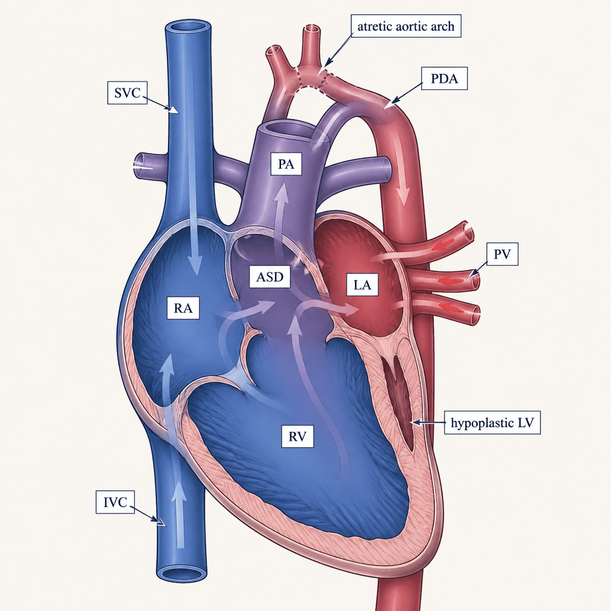
In a normal heart, two ventricles work in series: the right pumps blood to the lungs, and the left pumps oxygenated blood to the body. In single-ventricle anatomy, one ventricle is too small or malformed to do its job, so a single functional ventricle must be made to supply the body, while blood flow to the lungs is re-engineered surgically over three operations.
Stage 1 — The Norwood procedure (newborn period). Performed in the first days to weeks of life, the Norwood is among the highest-risk operations in all of congenital heart surgery. For HLHS it involves three key maneuvers: (1) reconstructing the aorta and systemic outflow so the single ventricle can pump to the body; (2) an atrial septectomy to allow unobstructed mixing of blood; and (3) creating a controlled source of pulmonary blood flow, either a modified Blalock-Taussig(-Thomas) shunt (MBTS) connecting an arm/head artery to the pulmonary artery, or a right-ventricle-to-pulmonary-artery (RV-PA, "Sano") shunt (Marino et al., AHA Scientific Statement, Circulation, 2018). After the Norwood, the child remains deeply vulnerable: oxygen saturations are intentionally low (typically high 70s to low 80s), and the fragile balance between blood flow to the lungs and to the body can collapse rapidly. A hybrid procedure — combining a catheter-based ductal stent with surgical pulmonary artery bands — is an alternative at some centers that defers the Norwood-type reconstruction (Ohye et al., Circulation, 2016).
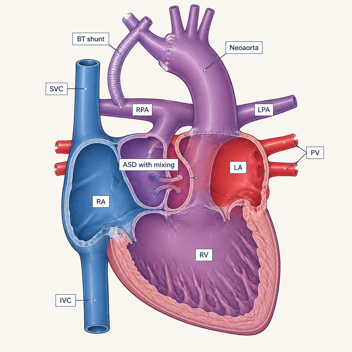
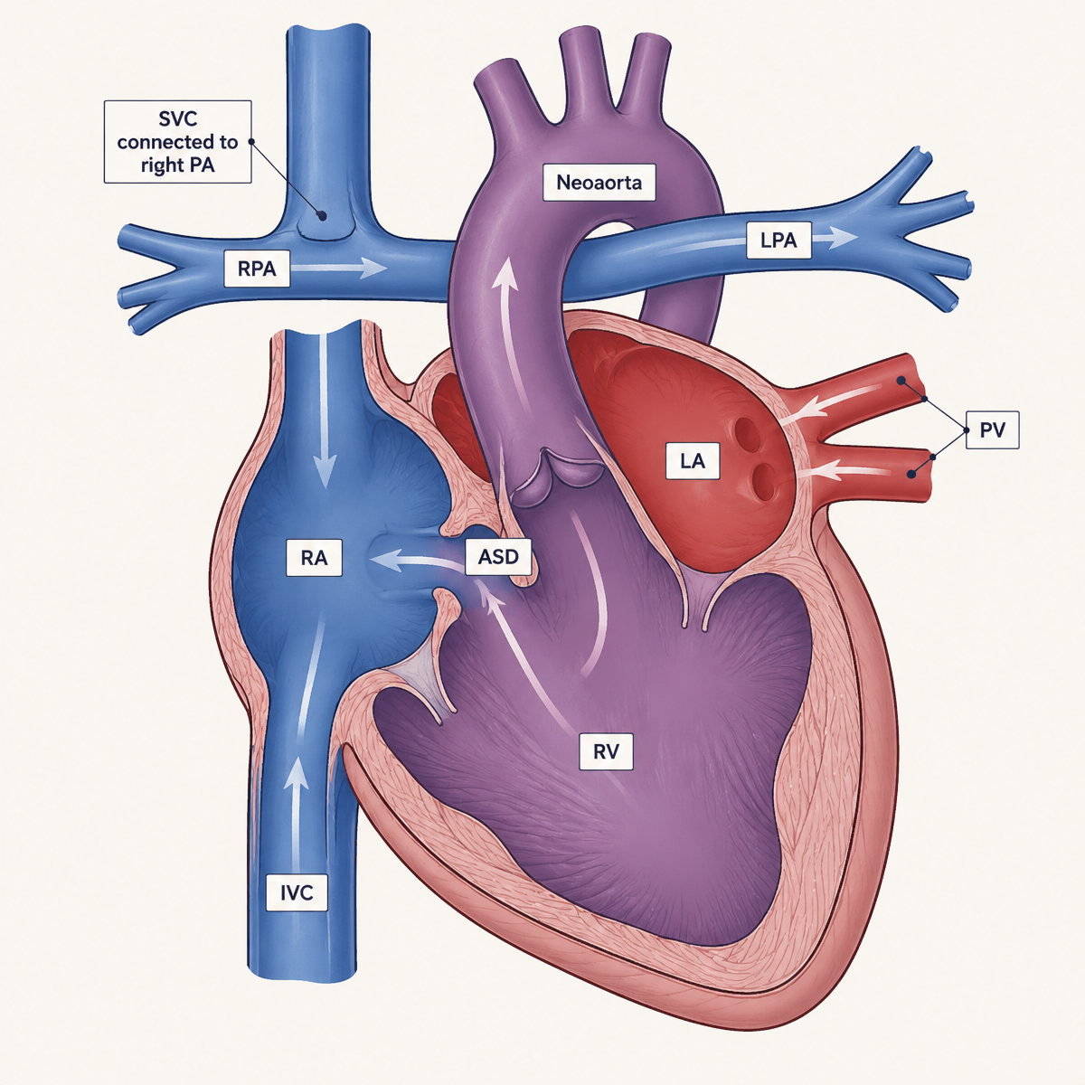
Stage 2 — The bidirectional Glenn / superior cavopulmonary anastomosis (about 3–6 months of age). The superior vena cava (the vein returning blood from the head and upper body) is connected directly to the pulmonary arteries, so that upper-body venous blood flows passively to the lungs. This "unloads" the single ventricle, reduces its volume burden, and removes the need for the earlier shunt. The Glenn is generally a much lower-risk operation than the Norwood, and the physiology it creates is considered the most stable of the three stages (Egidy Assenza et al., JAMA, 2026; Marino et al., AHA, 2018).
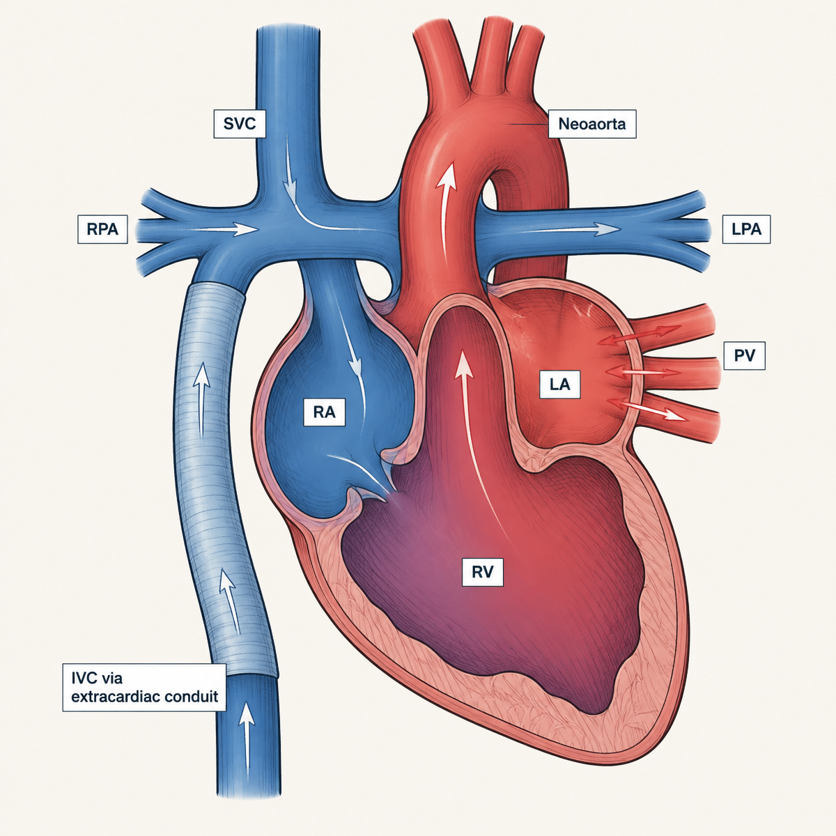
Stage 3 — The Fontan / total cavopulmonary connection (about 2–4 years of age). The inferior vena cava (returning blood from the lower body) is routed to the pulmonary arteries as well, usually via an extracardiac conduit or an intracardiac lateral tunnel, sometimes with a small "fenestration" (a pressure-relief hole). After the Fontan, essentially all systemic venous blood flows passively through the lungs without a pumping chamber, and oxygen saturations approach normal (Krishnamurthy et al., ACR Appropriateness Criteria, 2023; Rychik et al., AHA Scientific Statement, Circulation, 2019). The defining feature of the completed "Fontan circulation" is that pulmonary blood flow is driven not by a ventricle but by elevated systemic venous pressure and low pulmonary vascular resistance — a fragile arrangement with limited reserve.
Norwood vs. Glenn vs. Fontan at a Glance
The table below summarizes how the circulation changes at each stage. It is a conceptual comparison for attorneys reviewing records; individual surgical anatomy varies, and the operative reports — not general diagrams — define the circulation in a specific case.
Source of pulmonary blood flow
- Normal circulation
- The right ventricle pumps to the lungs
- After Norwood
- A systemic-to-pulmonary shunt (modified BT shunt) or RV-PA (Sano) conduit
- After Glenn
- SVC (upper-body) blood flows passively to the pulmonary arteries
- After Fontan
- SVC and IVC blood flow passively through the Fontan pathway
Role of the ventricle
- Normal circulation
- Two ventricles in series — one to the lungs, one to the body
- After Norwood
- The single ventricle supports both the body and, indirectly, the lungs
- After Glenn
- The single ventricle supports the body; upper-body return bypasses it
- After Fontan
- The single ventricle supports only the body
Systemic venous return
- Normal circulation
- All venous blood returns to the heart
- After Norwood
- All returns to the common atrium and ventricle, where it mixes
- After Glenn
- Upper-body (SVC) blood flows directly to the lungs; IVC blood still passes through the heart
- After Fontan
- All systemic venous blood is directed to the lungs without a subpulmonary ventricle
Degree of mixing
- Normal circulation
- None — the two circulations are separate
- After Norwood
- Complete mixing in the common atrium and ventricle
- After Glenn
- Partial — lower-body blood still mixes before reaching the aorta
- After Fontan
- Minimal in the Fontan pathway; oxygen saturations approach normal
Expected oxygenation
- Normal circulation
- Normal
- After Norwood
- Intentionally low (typically high 70s to low 80s)
- After Glenn
- Higher than after the Norwood, but still below normal
- After Fontan
- Near normal
Ventricular workload
- Normal circulation
- Two balanced pumps
- After Norwood
- High — the ventricle carries a large volume load
- After Glenn
- Reduced — the shunt is removed and upper-body flow bypasses the ventricle
- After Fontan
- Further reduced — no subpulmonary workload, but filling depends on venous pressure
Major physiologic vulnerabilities
- Normal circulation
- —
- After Norwood
- Imbalance between pulmonary and systemic flow; shunt obstruction; interstage fragility
- After Glenn
- Obstruction or thrombosis of the cavopulmonary connection; AV valve regurgitation; ventricular dysfunction
- After Fontan
- Fontan failure, venous congestion, arrhythmia, thrombosis, protein-losing enteropathy, plastic bronchitis, liver disease
Why dehydration or illness may matter
- Normal circulation
- Ordinary illness is usually tolerated
- After Norwood
- Dehydration, fever, or anemia can shift the flow balance abruptly
- After Glenn
- Passive flow depends on preload and pulmonary pressures; intercurrent illness can decompensate quickly
- After Fontan
- The circulation has limited reserve; dehydration or elevated pulmonary resistance can precipitate circulatory failure
Terminology commonly seen in records
- Normal circulation
- Sinus rhythm, biventricular circulation
- After Norwood
- Post-Norwood, MBTS, Sano conduit, neo-aorta, interstage
- After Glenn
- Bidirectional Glenn, SVC-PA anastomosis, cavopulmonary connection
- After Fontan
- Fontan circulation, extracardiac conduit, lateral tunnel, fenestration, PLE
| Normal circulation | After Norwood | After Glenn | After Fontan | |
|---|---|---|---|---|
| Source of pulmonary blood flow | The right ventricle pumps to the lungs | A systemic-to-pulmonary shunt (modified BT shunt) or RV-PA (Sano) conduit | SVC (upper-body) blood flows passively to the pulmonary arteries | SVC and IVC blood flow passively through the Fontan pathway |
| Role of the ventricle | Two ventricles in series — one to the lungs, one to the body | The single ventricle supports both the body and, indirectly, the lungs | The single ventricle supports the body; upper-body return bypasses it | The single ventricle supports only the body |
| Systemic venous return | All venous blood returns to the heart | All returns to the common atrium and ventricle, where it mixes | Upper-body (SVC) blood flows directly to the lungs; IVC blood still passes through the heart | All systemic venous blood is directed to the lungs without a subpulmonary ventricle |
| Degree of mixing | None — the two circulations are separate | Complete mixing in the common atrium and ventricle | Partial — lower-body blood still mixes before reaching the aorta | Minimal in the Fontan pathway; oxygen saturations approach normal |
| Expected oxygenation | Normal | Intentionally low (typically high 70s to low 80s) | Higher than after the Norwood, but still below normal | Near normal |
| Ventricular workload | Two balanced pumps | High — the ventricle carries a large volume load | Reduced — the shunt is removed and upper-body flow bypasses the ventricle | Further reduced — no subpulmonary workload, but filling depends on venous pressure |
| Major physiologic vulnerabilities | — | Imbalance between pulmonary and systemic flow; shunt obstruction; interstage fragility | Obstruction or thrombosis of the cavopulmonary connection; AV valve regurgitation; ventricular dysfunction | Fontan failure, venous congestion, arrhythmia, thrombosis, protein-losing enteropathy, plastic bronchitis, liver disease |
| Why dehydration or illness may matter | Ordinary illness is usually tolerated | Dehydration, fever, or anemia can shift the flow balance abruptly | Passive flow depends on preload and pulmonary pressures; intercurrent illness can decompensate quickly | The circulation has limited reserve; dehydration or elevated pulmonary resistance can precipitate circulatory failure |
| Terminology commonly seen in records | Sinus rhythm, biventricular circulation | Post-Norwood, MBTS, Sano conduit, neo-aorta, interstage | Bidirectional Glenn, SVC-PA anastomosis, cavopulmonary connection | Fontan circulation, extracardiac conduit, lateral tunnel, fenestration, PLE |
Why This Physiology Is Dangerous and Easy to Misjudge
Several features of single-ventricle physiology repeatedly surface in litigation because they defy the intuitions clinicians apply to otherwise healthy children:
- "Normal" saturation is low. A pre-Glenn or post-Norwood infant typically lives at oxygen saturations in the high 70s to low 80s. A saturation of 72% may be dangerously low for that child, or may be near baseline — the number is meaningless without the baseline. Treating such a child by healthy-infant standards (or, conversely, over-oxygenating them) can be harmful.
- Balance between lung and body blood flow is precarious. After the Norwood, too much blood flow to the lungs starves the body (and the coronary arteries), while too little causes profound hypoxia. Small changes — dehydration, fever, anemia — can tip this balance (Marino et al., AHA, 2018).
- Shunt obstruction is a true emergency. In a shunt-dependent infant, thrombosis or narrowing of the MBTS can cause rapid deterioration and arrest; restoring shunt patency (by catheterization or surgery), and sometimes ECMO as a bridge, is the recognized response (Lasa et al., PALS 2025 Guidelines, AHA/AAP).
- The interstage period is the highest-risk window. The interval between hospital discharge after the Norwood and the Glenn operation carries substantial mortality; in the Single Ventricle Reconstruction (SVR) trial, interstage mortality was about 12% overall and was higher with the MBTS than the RV-PA shunt, which is why home monitoring programs (daily weights and oxygen saturations with defined alert thresholds) became standard (Hill et al., JACC, 2026; Marino et al., AHA, 2018).
- Fontan patients decompensate through venous congestion, not just low blood pressure. Because the circulation depends on passive venous flow, these patients are exquisitely sensitive to dehydration, arrhythmia, and anything that raises pulmonary resistance; blood pressure can look reassuring while the circulation is failing.
Why the Stage Matters
Because the circulation is surgically reconstructed in stages, the same clinical finding can carry very different significance depending on where the child sits along the pathway. A given oxygen saturation, weight change, or feeding pattern may be near baseline for a post-Norwood infant, a warning sign in a post-Fontan child, or a defined red-flag threshold in an interstage home-monitoring program. Reviewing a single-ventricle case should therefore begin by establishing the child's stage of palliation and the documented baseline for that stage.
- Whether the child was preoperative, immediately postoperative, or remote from the last stage
- The expected baseline oxygen saturation for that stage and that specific child
- The specific anatomy and the specific operation performed — operative reports, not labels, define the circulation
- Ventricular and atrioventricular valve function on recent echocardiograms
- Respiratory status, hydration, and perfusion at presentation
- The intercurrent illness and its expected effect on the circulation at that stage
- Home-monitoring thresholds and prior documented clinical baselines, where available
No universal saturation threshold applies across all stages. Interpretation depends on the child's documented baseline and on the physiology created by the most recent operation.
Outcomes: What the Evidence Actually Shows
A central feature of these cases — for both plaintiff and defense — is that single-ventricle disease carries a high baseline risk of death and disability independent of the quality of care.
Norwood (Stage 1). Experienced centers now report hospital survival exceeding 90% for the Norwood, but mortality remains significant and highly center- and patient-dependent (Ohye et al., Circulation, 2016). In the contemporary SVR trial, 30-day and in-hospital mortality after the Norwood were roughly 11.5% and 16%, respectively (Hill et al., JACC, 2026). Reported first-stage mortality for HLHS is often cited at 15–20%, with wide variation across regions and programs (Silberberg et al., Pediatrics, 2025). Single-institution data show eight-year survival after the Norwood around 66%, with risk driven heavily by patient factors — prematurity, low weight, genetic or extracardiac anomalies, and the need for postoperative ECMO or unplanned reoperation (Alsoufi et al., Annals of Thoracic Surgery, 2015).
Glenn and Fontan (Stages 2 and 3). By contrast, the second and third operations have become comparatively low-risk, each commonly carrying a mortality risk of less than 1–2% in the current era (Silberberg et al., Pediatrics, 2025).
Long-term survival. More than 85% of children who complete the Fontan now survive into adulthood, and an estimated 70,000+ people worldwide live with a Fontan circulation (Glenn et al., Circulation, 2026). Patients operated on today may expect 30-year survival exceeding 80% (Rychik et al., AHA, 2019), and 20-year survival estimates range from 61% to 85% (Krishnamurthy et al., ACR, 2023). From a whole-cohort perspective, roughly half of neonates born with HLHS can be expected to reach age 15 without heart transplant, with survival strongly modified by associated cardiac and non-cardiac conditions (Silberberg et al., Pediatrics, 2025).
The burden of morbidity is high even among survivors. In the SVR trial long-term analysis, only about 12% of the original cohort remained free of major morbidity, and death was the single most common determinant of poor outcome, affecting roughly 39% of participants over follow-up (Hill et al., JACC, 2026). Cumulative Fontan-era complications include additional cardiac surgery, catheter interventions, arrhythmia requiring treatment, thrombosis, and protein-losing enteropathy, with about 10% dead or transplanted by early adulthood in one longitudinal cohort (Atz et al., JACC, 2017).
Late Fontan complications. The Fontan circulation predisposes to arrhythmia, thromboembolism, progressive ventricular and valve dysfunction, liver fibrosis, renal dysfunction, protein-losing enteropathy, plastic bronchitis, and neurocognitive and psychological challenges. The risk of "Fontan circulatory failure" approaches 40% at 35 years of follow-up (Egidy Assenza et al., JAMA, 2026; Rychik et al., AHA, 2019). Quality-of-life studies show children and adults with Fontan physiology report lower health-related quality of life than population norms, with psychological factors (anxiety, depression) strongly influencing outcomes (Marshall et al., Journal of Pediatrics, 2024).
These figures matter legally because they define the backdrop against which any alleged deviation must be measured: a poor outcome in a single-ventricle child is frequently the expected natural history of the disease, not evidence of negligence.
Standard of Care and Surveillance
There is no single "standard of care" document covering every decision in single-ventricle management, and attorneys should be cautious about experts who overstate the firmness of the evidence. The American Heart Association's 2019 scientific statement on Fontan circulation expressly notes that, by strict evidence-grading criteria, there is insufficient evidence to mandate specific surveillance tests or intervals; instead it offers a consensus-based surveillance "toolkit" and emphasizes individualization and care by clinicians experienced with this physiology (Rychik et al., AHA, 2019). Surveillance practices vary considerably across centers (Di Maria et al., Pediatric Cardiology, 2019; Onorato et al., Pediatric Cardiology, 2025).
Recognized elements of contemporary care — while acknowledging the limits above — commonly include:
- Interstage home monitoring after the Norwood (daily oxygen saturation and weight with defined "red-flag" thresholds prompting contact), developed in response to high interstage mortality (Marino et al., AHA, 2018).
- Care coordinated with, or delivered by, a pediatric cardiology/single-ventricle program. When an interstage or Fontan patient presents acutely, early involvement of pediatric cardiology is widely expected; these patients are explicitly recognized as requiring management by providers familiar with the physiology (Rychik et al., AHA, 2019; Lasa et al., PALS 2025).
- Emergency recognition of shunt obstruction and low-cardiac-output states. The 2025 PALS guidelines address resuscitation specific to single-ventricle patients, including measures to restore shunt patency and the role of ECMO; notably, clinical practice favors earlier recognition of the postoperative low-output state and elective ECMO before arrest, as outcomes after cardiac arrest in this population are poor (survival to discharge roughly 32–57% in reported series) (Lasa et al., PALS 2025).
- Stage-appropriate imaging surveillance (echocardiography as the mainstay; cardiac MRI, catheterization, and other studies when transthoracic imaging is inadequate), with frequency tailored to stage (Krishnamurthy et al., ACR, 2023).
When evaluating a case, the key question is rarely whether a textbook rule was broken, but whether the clinicians recognized that this was a single-ventricle patient, obtained and interpreted the child's baseline physiology, and escalated appropriately.
Where Litigation Typically Arises (Targets for Plaintiffs)
- Failure to recognize or act on the interstage patient. The post-Norwood, pre-Glenn infant who presents with feeding problems, lethargy, respiratory symptoms, or a saturation change, and is discharged without cardiology involvement or shunt evaluation.
- Failure to appreciate baseline physiology. Treating low baseline saturations as either falsely reassuring or falsely alarming; failing to document and compare against the child's known baseline.
- Delayed diagnosis of shunt obstruction or a low-cardiac-output state. Missing a surgical/catheterization emergency, or failing to deploy ECMO before arrest in a deteriorating postoperative patient.
- Surgical and perioperative complications. Technical imperfections requiring reoperation are associated with failure to progress through the stages and with reduced survival (Alsoufi et al., Annals of Thoracic Surgery, 2015); allegations may involve the index operation, postoperative ICU management, or recognition of complications.
- Inadequate interstage monitoring or home-monitoring failures. Absence of a monitoring program, or failure to respond to breach of alert thresholds.
- Missed or delayed late Fontan complications. Arrhythmia, thrombosis/stroke, protein-losing enteropathy, plastic bronchitis, liver disease, or progressive ventricular failure that is not surveilled for or acted upon.
- Communication and handoff failures among emergency physicians, hospitalists, cardiologists, and surgeons — a recurring theme across congenital-heart litigation.
- Documentation deficiencies, particularly incomplete vital signs, absent perfusion assessment, and no recorded baseline comparison.
Common Defenses
- Natural history of a high-risk disease. The outcome data above establish that death and major morbidity are common even with optimal care; a bad outcome is often the expected course of the lesion, not a result of any deviation.
- Patient-specific, non-modifiable risk factors. Prematurity, low birth weight, genetic syndromes, extracardiac anomalies, ventricular dysfunction, and valve regurgitation independently drive mortality (Alsoufi et al., 2015; Marino et al., AHA, 2018).
- Limits of the evidence base. For many surveillance and management decisions, guidelines are consensus-based and expressly decline to mandate specific tests or intervals, leaving substantial room for legitimate physician judgment (Rychik et al., AHA, 2019).
- Causation defenses. Even where a deviation is conceded, the defense may show that earlier intervention would not, more likely than not, have changed the outcome — for example, that arrest or circulatory failure was already inevitable, or that the complication was not reasonably foreseeable or preventable.
- Appropriate and timely escalation. Documentation that cardiology was involved, baseline physiology was considered, and recognized emergency measures were undertaken.
How Causation Is Analyzed
Causation is frequently the decisive issue, precisely because the baseline risk is so high. The analysis typically asks:
- Would earlier or different action, more likely than not, have changed the outcome? This requires distinguishing probability from mere possibility, and situating the specific child within the known outcome data.
- Was the injury the natural progression of the lesion, an unavoidable complication, or the result of a deviation? Interstage death, shunt thrombosis, and Fontan circulatory failure can each occur despite excellent care.
- Was the complication foreseeable, and was there a realistic window to intervene? For shunt-dependent infants and deteriorating postoperative patients, the literature supports earlier recognition and elective ECMO as potentially outcome-changing, but post-arrest survival is poor (Lasa et al., PALS 2025).
- Did pre-existing, non-cardiac conditions independently drive the outcome? Genetic and extracardiac anomalies substantially lower survival regardless of cardiac care (Silberberg et al., Pediatrics, 2025).
Long-Term Damages Considerations
Damages analysis in surviving patients must account for the lifelong, multisystem nature of the Fontan circulation: recurrent surgeries and catheterizations, arrhythmia management, anticoagulation, risk of thromboembolic stroke, protein-losing enteropathy and plastic bronchitis, Fontan-associated liver disease, renal dysfunction, exercise limitation, neurodevelopmental and psychological impairment, potential need for heart transplantation, and reduced health-related quality of life (Egidy Assenza et al., JAMA, 2026; Rychik et al., AHA, 2019; Atz et al., JACC, 2017; Marshall et al., Journal of Pediatrics, 2024). Life-care planning in these cases is complex and should involve clinicians familiar with the trajectory of single-ventricle survivors.
Medical Records That Matter
- Fetal echocardiography and prenatal counseling records
- Operative notes for each stage (Norwood/hybrid, Glenn, Fontan) and any reoperations
- Cardiac ICU documentation, including hemodynamics, ventilation, and ECMO records
- Interstage home-monitoring logs (daily weights, saturations, alert thresholds and responses)
- Serial echocardiograms, cardiac catheterizations, and cardiac MRI
- Pediatric cardiology clinic notes and surveillance testing
- Emergency department and urgent-care records, with complete vital signs, perfusion assessment, and documented baseline saturation
- Growth, feeding, and developmental history
- Communication/handoff records among emergency, hospitalist, cardiology, and surgical teams
Isolated records rarely tell the story; the longitudinal trajectory across stages is usually essential.
The Value of Pediatric Cardiology Expertise
Single-ventricle care spans fetal diagnosis, neonatal surgery, intensive care, interstage home monitoring, interventional catheterization, and decades of multisystem follow-up. The physiology is stage-dependent and counterintuitive, "normal" values differ from those in healthy children, and the evidence base is in important respects consensus-driven rather than prescriptive. A qualified pediatric cardiology expert can interpret the anatomy and imaging, place the specific child within the correct outcome data, distinguish deviation from natural history, and explain causation in terms a jury can follow — while an unqualified expert may overstate the firmness of "standards" that the literature itself describes as uncertain.
Terms Attorneys May See in the Record
- Single ventricle — A group of congenital heart defects in which only one ventricle is adequate to support the circulation; a physiologic description rather than one specific anatomy.
- HLHS (hypoplastic left heart syndrome) — The most common single-ventricle lesion, in which the left-sided structures (left ventricle, aortic valve, and aorta) are underdeveloped.
- Norwood — The Stage 1 neonatal operation (or a hybrid alternative) that reconstructs the systemic outflow, ensures unobstructed atrial mixing, and creates a controlled source of pulmonary blood flow.
- Neo-aorta — The reconstructed systemic outflow tract created during the Norwood, arising from the single ventricle.
- BT shunt (modified Blalock-Taussig-Thomas shunt, MBTS) — A small synthetic tube from a branch of the aortic arch to a pulmonary artery that supplies pulmonary blood flow after the Norwood.
- Sano conduit (RV-PA shunt) — A conduit from the right ventricle directly to the pulmonary artery, used at some centers as the Stage 1 source of pulmonary blood flow.
- Hybrid procedure — A Stage 1 alternative combining a catheter-based ductal stent with surgical pulmonary artery bands, deferring full Norwood-type reconstruction.
- Interstage — The period between discharge after the Norwood and the Glenn operation, during which many programs use structured home monitoring.
- Glenn (bidirectional Glenn, superior cavopulmonary anastomosis) — Stage 2: the SVC is connected directly to the pulmonary arteries so upper-body blood flows passively to the lungs.
- Cavopulmonary connection — The general term for surgically connecting systemic veins to the pulmonary arteries (encompassing the Glenn and the Fontan).
- Fontan (total cavopulmonary connection) — Stage 3: inferior vena caval blood is also routed to the pulmonary arteries, completing the passive pulmonary circulation.
- Fontan conduit — The pathway (an extracardiac conduit or an intracardiac lateral tunnel) that carries IVC blood to the pulmonary arteries.
- Fenestration — A small, intentional pressure-relief opening between the Fontan pathway and the atrium, created in some patients and later closed in many.
- Systemic ventricle — The single functional pumping chamber that supports the body circulation.
- Pulmonary vascular resistance (PVR) — The resistance to blood flow within the lung vessels; a key determinant of whether passive Fontan flow is feasible.
- AV valve regurgitation — Leaking of the valve or valves between the atrium and the single ventricle, which increases the ventricle's volume load.
- Ventricular dysfunction — Impaired contraction or relaxation of the single ventricle.
- Cyanosis — Bluish discoloration of the skin or lips caused by low blood oxygen; expected to some degree at the earlier stages.
- Oxygen saturation (SpO2) — The percentage of hemoglobin carrying oxygen; in these children it must be interpreted against the stage-specific documented baseline.
- Qp:Qs — The ratio of pulmonary blood flow to systemic blood flow; a way of describing the balance between the two circuits.
Key Takeaways for Attorneys
- Single-ventricle disease is managed, not cured, through three staged operations: Norwood (newborn), Glenn (3–6 months), and Fontan (2–4 years).
- The Norwood and the interstage period are the highest-risk windows; the Glenn and Fontan are comparatively low-risk operations.
- "Normal" oxygen saturation is low in these children; the number is meaningless without the documented baseline.
- Shunt obstruction and low-cardiac-output states are emergencies; recognized care favors early escalation, cardiology involvement, and elective ECMO before arrest.
- Baseline mortality and morbidity are high even with flawless care — a poor outcome is frequently the disease's natural history, which is central to both liability and causation.
- Key guidelines (notably the AHA Fontan statement) are often consensus-based and decline to mandate specific tests or intervals, leaving room for legitimate physician judgment.
- Causation turns on probability, not possibility, and on whether a realistic window to change the outcome existed.
- Documentation gaps — incomplete vitals, absent perfusion assessment, no baseline comparison, and handoff failures — are recurring targets.
- Damages are lifelong and multisystem, requiring expert life-care planning.
- Credible evaluation requires a pediatric cardiology expert familiar with stage-specific physiology and the actual outcome literature.
References
- Glenn et al., Circulation, 2026.
- Ohye et al., Circulation, 2016.
- Silberberg et al., Pediatrics, 2025.
- Marino et al., AHA Scientific Statement, Circulation, 2018.
- Egidy Assenza et al., JAMA, 2026.
- Krishnamurthy et al., ACR Appropriateness Criteria, 2023.
- Rychik et al., AHA Scientific Statement, Circulation, 2019.
- Lasa et al., PALS 2025 Guidelines, AHA/AAP.
- Hill et al., JACC, 2026.
- Alsoufi et al., Annals of Thoracic Surgery, 2015.
- Atz et al., JACC, 2017.
- Marshall et al., Journal of Pediatrics, 2024.
- Di Maria et al., Pediatric Cardiology, 2019.
- Onorato et al., Pediatric Cardiology, 2025.
Educational Disclaimer
This article is provided for educational purposes only and does not constitute legal or medical advice. Every case is unique and should be evaluated on its individual facts and complete medical records.
About the Author
Asif Masood, MD, MSc is double board-certified in General Pediatrics and Pediatric Cardiology. Through The Verdict MD, he provides expert witness services in pediatric and congenital cardiology and general pediatrics, as well as independent medical consulting for attorneys, law firms, insurers, and healthcare organizations nationwide.