Pediatric & Congenital Cardiology
Complete Atrioventricular Septal Defect (AV Canal): A Medical-Legal Guide for Attorneys
- Published
- November 9, 2026
- Last Reviewed
- November 9, 2026
- Author
- Asif Masood, MD, MSc
- Reading Time
- 16 min
- Category
- Pediatric & Congenital Cardiology
This guide reviews complete atrioventricular septal defect for attorneys evaluating prenatal and newborn diagnosis, recognition of heart failure, surgical timing and strategy, complications, and medical causation.
Introduction
Complete atrioventricular canal defect, also called complete atrioventricular septal defect (CAVC or complete AVSD), is a major congenital heart defect. It makes up about 4–5% of all congenital heart disease and is the most common heart defect in children with Down syndrome (trisomy 21) (Gurvitz et al., Journal of the American College of Cardiology, 2026; Dimopoulos et al., Circulation, 2023). With surgery at the right time, most children do well over the long term. The defect is unforgiving of delay, though. Without repair, the lungs can be permanently damaged by excess blood flow and high pressure. Once that damage sets in, repair may no longer be possible.
For attorneys, CAVC cases tend to involve one or more of four themes:
- missed prenatal or newborn diagnosis
- missed or late recognition of heart failure and pulmonary hypertension in infancy
- surgical decisions, especially in the "unbalanced" form, where a two-ventricle repair and a single-ventricle pathway are both possible
- postoperative complications such as valve leakage, heart block, and need for reoperation
This article explains the medicine behind each theme. It is educational and does not express an opinion on liability in any case. Medical guidelines describe evidence-based practice. The legal standard of care depends on the jurisdiction and is decided by the courts.
Hypothetical Case
The following case is fictional and is presented for educational purposes only.
Baby J, a girl, is born at term after a pregnancy with a second-trimester anatomy scan reported as "limited cardiac views due to fetal position; otherwise normal." No follow-up scan or fetal echocardiogram is ordered. At birth, features of Down syndrome are noted, and genetic testing is sent. Her newborn pulse oximetry screen passes, at 96% in both the hand and the foot. No murmur is heard, and she goes home on day 2. The discharge summary says, "Chromosome results pending; follow up with PCP."
At her 2-week visit, the pediatrician notes that the karyotype has confirmed trisomy 21. No echocardiogram has been done. The record lists "cardiology referral," but no order or appointment appears. At 6 and 9 weeks, the parents report rapid breathing, sweating during feeds, and long feeding times. Her weight drops from the 25th to below the 3rd percentile. A soft murmur is documented as "likely flow murmur."
At 4 months, she is seen in the emergency department with respiratory distress. An echocardiogram shows a complete AVSD with moderate valve leakage, and the defect is balanced. Pulmonary pressures are close to systemic levels. Cardiac catheterization shows elevated pulmonary vascular resistance that partly responds to oxygen and nitric oxide. She undergoes repair at 5 months. Afterward she has pulmonary hypertensive crises, a long ICU stay, and continued need for pulmonary hypertension medicine.
The case raises several questions an expert would examine:
- Should the limited prenatal views have led to further imaging?
- Was the postnatal echocardiogram that is recommended for every infant with Down syndrome ordered and completed?
- Were the signs of heart failure recognized?
- Was referral timely?
- Did any delay change her lung outcome, given that infants with Down syndrome can develop pulmonary vascular disease early even with good care?
Understanding the Defect
Anatomy. The center of the heart normally has a wall between the upper chambers (atria), a wall between the lower chambers (ventricles), and two separate inlet valves (mitral and tricuspid). In CAVC, the tissue at this center, called the endocardial cushions, fails to develop. The result is three linked abnormalities:
- a hole between the atria (a primum atrial septal defect)
- a hole between the ventricles (an inlet ventricular septal defect)
- a single common atrioventricular valve in place of two separate valves, often leaky
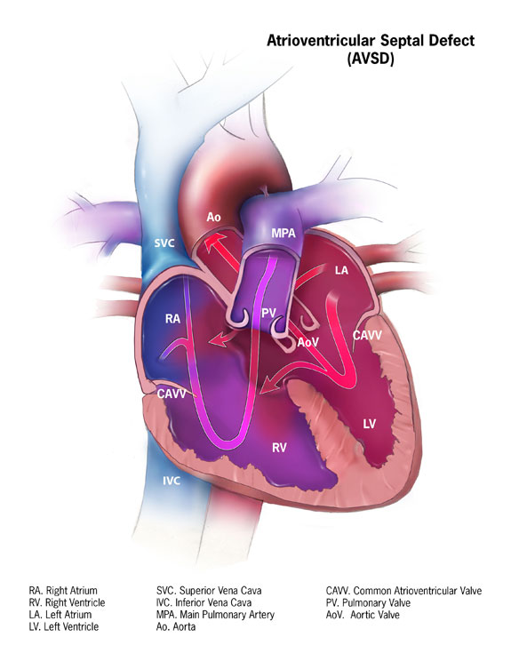
The 2025 ACC/AHA adult congenital heart disease guideline separates three forms. In partial AVSD there is only an atrial-level hole, often with a cleft in the left valve. In transitional AVSD the ventricular hole is small and restrictive. In complete AVSD there are large holes at both the atrial and ventricular levels (Gurvitz et al., Journal of the American College of Cardiology, 2026). This article covers the complete form.
Physiology. Pressure in the left side of the heart is higher than in the right. Blood therefore shunts from left to right through the large defects and floods the lungs. Over the first weeks of life, the normal fall in lung resistance increases this shunt. Infants develop congestive heart failure, which shows up as fast breathing, sweating and tiring with feeds, poor weight gain, and frequent respiratory infections. A leaky common valve makes this worse.
Over time, high flow and pressure damage the small lung arteries. This is pulmonary vascular disease. If it becomes irreversible, it can end in Eisenmenger physiology, where the shunt reverses, the child becomes cyanotic, and surgical repair is no longer an option.
Balanced versus unbalanced. In a balanced CAVC, the common valve sits evenly over two well-sized ventricles. In an unbalanced CAVC, the valve sits mostly over one ventricle, and the other ventricle is underdeveloped. Most often the right ventricle is dominant and the left is small. Less often the left is dominant.
Clinicians measure balance with echocardiographic indices. The best known is the atrioventricular valve index (AVVI), which is the area of the left part of the valve divided by the total valve area. A Congenital Heart Surgeons' Society study of 356 infants found the following (Jegatheeswaran et al., Circulation, 2010):
- AVVI between 0.4 and 0.6: balanced defect, and these children usually had a two-ventricle repair
- AVVI below 0.19: all children had a single-ventricle pathway
- AVVI from 0.19 to 0.39: the "gray zone," where surgeons chose different strategies and deaths were disproportionately frequent
Other measures include ventricular size ratios, inflow angles, and cardiac MRI. Accepted cutoffs for deciding between one- and two-ventricle repair have not been established (Deal et al., Pediatric Cardiac Surgery, 2023; Schleiger et al., Pediatric Cardiology, 2021).
Associations. About half of infants with Down syndrome have congenital heart disease, and AVSD is the most common lesion (Dimopoulos et al., Circulation, 2023). Down syndrome is also linked to a high rate of unbalanced AVSD. Unbalanced AVSD is common in heterotaxy syndromes as well, which often come with additional heart and organ abnormalities. In one surgical series of unbalanced defects, about half of the children had heterotaxy (Nathan et al., European Journal of Cardio-Thoracic Surgery, 2017).
Prenatal Diagnosis
Unlike transposition of the great arteries, CAVC usually changes the standard four-chamber view on a routine anatomy scan. The center of the heart, where the walls and valves normally meet, looks abnormal. For this reason, CAVC is generally one of the more detectable major defects before birth. Detection still depends on image quality, fetal position, maternal body habitus, gestational age, and the skill of the operator.
Common medicolegal questions include:
- Were adequate cardiac views obtained?
- Were "limited" or "suboptimal" views followed by a repeat scan or a fetal echocardiogram?
- Did abnormal genetic screening (cell-free DNA, nuchal translucency, or other markers of trisomy 21) lead to fetal echocardiography?
- Was the diagnosis communicated so that delivery could be planned?
A missed prenatal diagnosis is not always substandard care. Unlike d-TGA, though, prenatal detection of CAVC rarely changes the newborn's immediate survival. The infant is usually stable at birth. The harms that matter tend to come later, from delayed recognition after birth. Causation arguments based only on a missed prenatal diagnosis are therefore often limited. The exceptions include lost reproductive decision-making (a separate legal theory in some jurisdictions) and cases where the missed diagnosis led directly to a missed newborn diagnosis.
Postnatal Recognition
Why newborns may look well. At birth, lung resistance is still high, so the left-to-right shunt is small. Many infants with CAVC have no symptoms and may have no loud murmur in the first days. Oxygen saturations are usually near normal. Pulse oximetry screening therefore often does not detect CAVC. A passing screen does not rule out the defect.
Clues. Findings that should prompt evaluation include:
- known or suspected Down syndrome
- a murmur, especially one that appears or gets louder over the first weeks
- signs of heart failure: fast breathing, sweating or tiring with feeds, poor weight gain, liver enlargement
- frequent or severe respiratory infections
- an ECG showing a "superior" (left) axis, which is characteristic of AVSD because the conduction system is displaced (Gurvitz et al., Journal of the American College of Cardiology, 2026)
Down syndrome is a key screening point. The American Academy of Pediatrics recommends that every infant with Down syndrome have an echocardiogram read by a pediatric cardiologist, whether or not a fetal echocardiogram was done (Lin et al., American Journal of Medical Genetics Part C, 2020). Missing this step, or ordering it and failing to complete it, is one of the most frequently examined issues in CAVC cases.
Diagnostic Evaluation and Management
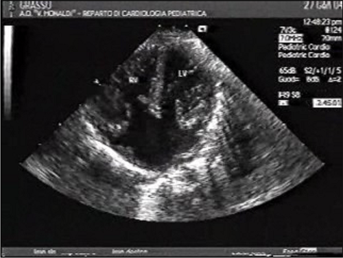
Echocardiography confirms the diagnosis. It defines the type of defect, the valve anatomy and leakage, ventricular balance, associated lesions, and estimated lung pressures. In unbalanced or borderline cases, cardiac MRI can help judge whether the smaller ventricle is big enough for a two-ventricle repair (Deal et al., Pediatric Cardiac Surgery, 2023). Cardiac catheterization is used when lung vascular resistance is uncertain, particularly in older infants or those with Down syndrome.
Medical therapy until surgery. This includes diuretics, afterload reduction, fortified feeds, and sometimes tube feeding to support growth (Sachdeva et al., Journal of the American College of Cardiology, 2026). Medical therapy is a bridge. It does not replace timely repair.
Timing of repair. The 2026 ACC Concise Clinical Guidance recommends considering surgical repair for all patients with complete AVSD between 3 and 6 months of age. It stresses that repair in this window is especially important in trisomy 21 and other pulmonary hypertension risk factors, to prevent irreversible pulmonary vascular disease (Sachdeva et al., Journal of the American College of Cardiology, 2026). Some infants develop heart failure that cannot be controlled and need earlier surgery. In one propensity-matched study of infants younger than 3 months, primary complete repair had 20-year survival of 94.2%, compared with 58.4% for staged repair after pulmonary artery banding (Buratto et al., Journal of Thoracic and Cardiovascular Surgery, 2021).
Balanced CAVC. The standard operation is a two-ventricle (biventricular) repair. The surgeon closes the atrial and ventricular holes with patches and divides the common valve into two working valves, usually closing the left valve cleft. Perioperative mortality is low. Long-term survival after complete AVSD repair is reported at 91–92% at 15 years (Gurvitz et al., Journal of the American College of Cardiology, 2026).
Unbalanced CAVC. There are three main strategies:
- Biventricular repair, if the smaller ventricle and its valve are adequate.
- Single-ventricle palliation. This is a series of operations (pulmonary artery banding or a shunt, then a Glenn, then a Fontan) that uses one ventricle to pump blood to the body.
- Staged biventricular recruitment or conversion. The surgeon starts on a single-ventricle path while trying to grow the smaller ventricle, then converts to a two-ventricle circulation.
Mortality with single-ventricle palliation in unbalanced AVSD is substantial. A biventricular strategy is generally favored when it is feasible (Schleiger et al., Pediatric Cardiology, 2021). In one tertiary series of 212 patients, children who underwent biventricular conversion or recruitment had transplant-free survival similar to those who had primary biventricular repair, and better than those who stayed on the single-ventricle path. Reinterventions were common in every group: 51.9% overall, with 18.9% mortality across the cohort (Nathan et al., European Journal of Cardio-Thoracic Surgery, 2017).
For children with Down syndrome on a single-ventricle path, protecting the lungs with a timely pulmonary artery band is essential. Early pulmonary vascular disease can make a later Glenn or Fontan impossible. Expert consensus states that Down syndrome by itself should not determine the choice of surgery (Dimopoulos et al., Circulation, 2023).
Complications After Repair
These complications are known risks of CAVC repair. Their occurrence does not by itself show substandard care.
- Left AV valve regurgitation. This is the most common reason for reoperation. Abnormal valve anatomy, such as a single papillary muscle or a double-orifice valve, raises the risk (Gurvitz et al., Journal of the American College of Cardiology, 2026; Deal et al., Pediatric Cardiac Surgery, 2023).
- Left ventricular outflow tract obstruction. This is the second most common reason for reoperation (Gurvitz et al., Journal of the American College of Cardiology, 2026).
- Heart block. The conduction system runs near the edge of the ventricular hole, so it is at risk during surgery. Complete heart block may need a permanent pacemaker. It can also appear late, especially in patients who had conduction problems after surgery (Gurvitz et al., Journal of the American College of Cardiology, 2026).
- Residual shunts, arrhythmias, and pulmonary hypertensive crises, the last especially with late repair or Down syndrome.
- Reintervention. About 15% of patients with partial or complete AVSD need surgical reintervention (Sachdeva et al., Journal of the American College of Cardiology, 2026).
Lifelong surveillance is required. Patients without significant residual lesions are generally seen every 1 to 3 years after the first postoperative year. Those with significant residual lesions are seen every 3 to 12 months (Sachdeva et al., Journal of the American College of Cardiology, 2026).
Targets for Litigation: Issues Commonly Examined
The following are areas experts commonly review. Whether any of them amounts to a breach of the legal standard of care depends on the facts and the jurisdiction.
- Prenatal imaging. This covers failure to get adequate four-chamber views, failure to follow up "limited" views, failure to refer for fetal echocardiography after abnormal aneuploidy screening, and failure to tell the family about findings.
- Missed Down syndrome cardiac screening. This covers no postnatal echocardiogram, an echocardiogram ordered but never completed, or results never followed up. Handoff failures between the birth hospital and the primary care practice are frequent.
- Reliance on a normal newborn exam or pulse oximetry screen. CAVC often does not lower oxygen saturation at birth. Using a passed screen to rule out heart disease in an infant with Down syndrome or a new murmur is a common issue.
- Failure to recognize heart failure. This covers fast breathing, sweating with feeds, and falling growth percentiles attributed to reflux, feeding technique, or "Down syndrome hypotonia," and new murmurs called innocent without evaluation.
- Delayed referral or surgery. The question is whether repair happened within the recommended 3–6 month window, and if not, why. Possible reasons include system delays, waitlists, intercurrent illness, family factors, or a clinical decision.
- Pulmonary vascular disease. Late repair may leave the child with irreversible pulmonary hypertension, a defect that can no longer be operated on, or a failed single-ventricle path. This is often the central damages issue.
- Unbalanced CAVC decision-making. Issues include whether the imaging (echocardiography, MRI) and multidisciplinary review were adequate before choosing one- versus two-ventricle repair, whether the family was told about the alternatives (informed consent), and whether a pulmonary artery band was placed in time to protect the lungs. Because no validated cutoffs exist in the gray zone, reasonable surgeons may disagree. Disagreement alone is not evidence of negligence.
- Intraoperative and postoperative care. Issues include recognition and management of heart block (temporary pacing, timing of a permanent pacemaker), residual valve leakage found on intraoperative transesophageal echocardiography and whether it was addressed, management of pulmonary hypertensive crises, and timely recognition of low cardiac output.
- Follow-up. Issues include loss to follow-up after repair, missed progressive valve leakage or outflow obstruction, and failure to monitor for late heart block.
- Documentation and communication. Issues include unrecorded growth trends, missing referral orders, unclear discharge instructions, and incomplete informed consent records.
Medical Causation
Causation analysis in CAVC is usually about time and the lungs. Typical questions include:
- When would timely diagnosis have occurred?
- When would repair have been done?
- What were the lung pressures and resistance at each point?
- Would earlier repair, more likely than not, have avoided the specific harm?
Specific harms might include irreversible pulmonary hypertension, an extended ICU course, or loss of candidacy for repair.
Several factors make causation hard to establish:
- Infants with Down syndrome can develop pulmonary arterial hypertension within the first year even with good care. The reported incidence ranges from 6% to 37.5%, and contributing causes include lung developmental abnormalities and airway disease (Dimopoulos et al., Circulation, 2023).
- Valve anatomy, not timing, often drives later valve leakage and reoperation.
- In unbalanced CAVC, the anatomy itself sets much of the risk. Mortality is concentrated in the gray zone no matter which strategy is chosen (Jegatheeswaran et al., Circulation, 2010).
- Neurodevelopmental outcomes in children with Down syndrome reflect the syndrome itself. Experts must separate any added effect of heart failure, malnutrition, or surgical complications from the baseline.
Opinions generally must be stated to a reasonable degree of medical probability, not mere possibility. The expert must explain the specific mechanism linking the delay to the injury.
Long-Term Outcomes
After a timely biventricular repair of balanced CAVC, most children survive into adulthood with good function. Long-term survival is reported at 91–92% at 15 years (Gurvitz et al., Journal of the American College of Cardiology, 2026). These children still need lifelong cardiology follow-up, and a meaningful minority need reoperation, mostly for left valve leakage or outflow obstruction.
Outcomes after single-ventricle palliation are more guarded. Patients face Fontan-related complications over a lifetime, including liver disease, arrhythmias, and eventual circulatory failure. Mortality is higher in people with Down syndrome and single-ventricle physiology (Dimopoulos et al., Circulation, 2023). Irreversible pulmonary vascular disease from late repair carries the worst prognosis, with lifelong pulmonary hypertension therapy, reduced exercise capacity, and shortened life expectancy.
Medical Records That Often Matter
- Prenatal ultrasound reports and images, aneuploidy screening results, and fetal echocardiogram referrals
- Nursery records, newborn exam findings, and pulse oximetry values
- Genetic test results and the date they were communicated
- Primary care growth charts, feeding histories, and murmur documentation
- Referral orders, scheduling records, and missed-appointment notices
- Echocardiograms (images, not only reports) and cardiac MRI and catheterization data, including lung pressures and resistance
- Surgical conference notes, informed consent documents, operative reports, and intraoperative transesophageal echocardiograms
- ICU records, pacing and rhythm strips, and pulmonary hypertension management
- Long-term cardiology follow-up and reintervention records
Common Misconceptions
- "A passed pulse oximetry screen rules out serious heart disease." CAVC often causes little or no drop in oxygen saturation at birth.
- "No murmur in the nursery means a normal heart." The murmur may be faint or absent until lung resistance falls.
- "Poor feeding is expected in Down syndrome." Low muscle tone affects feeding, but sweating, fast breathing, and falling growth percentiles suggest heart failure.
- "Reoperation means the first surgery was done poorly." Reoperation for valve leakage or outflow obstruction is a known part of the natural history after repair.
- "Choosing a single-ventricle path for an unbalanced defect was obviously wrong." In the gray zone, strategy is a matter of individualized expert judgment without validated cutoffs.
- "Pulmonary hypertension always means repair was late." In Down syndrome, it can develop early despite appropriate care.
The Value of Pediatric Cardiology Expertise
CAVC cases call for expertise in several areas:
- fetal and postnatal cardiac imaging
- assessment of ventricular balance
- pulmonary vascular physiology and catheterization data
- surgical decision-making and complications
- the specific cardiovascular course in Down syndrome and heterotaxy
An independent review of the actual images and hemodynamic data is often essential. Reports alone may not show ventricular balance, valve anatomy, or lung pressure trends.
Key Takeaways for Attorneys
- Complete AVSD involves holes between the atria and the ventricles plus a single, often leaky, common valve. It is strongly associated with Down syndrome.
- Newborns with CAVC often look well and pass pulse oximetry screening. Heart failure typically develops over weeks.
- The AAP recommends an echocardiogram for every infant with Down syndrome. Gaps in this screening are a frequent issue.
- The ACC recommends considering repair between 3 and 6 months of age to prevent irreversible pulmonary vascular disease, a recommendation that is especially important in Down syndrome.
- Balanced CAVC is repaired with a two-ventricle operation, with long-term survival of about 91–92% at 15 years.
- Unbalanced CAVC includes a gray zone where reasonable surgeons may choose different strategies. Disagreement is not proof of negligence, but the adequacy of evaluation and informed consent can be reviewed.
- Left valve leakage, outflow obstruction, and heart block (including late-onset block) are recognized complications. Their occurrence alone does not establish substandard care.
- Causation often turns on lung pressure and resistance trends over time, considered against the baseline risk of early pulmonary hypertension in Down syndrome.
- Review of the actual imaging, catheterization data, and operative findings is often essential.
- Medical guidelines inform the analysis, but the legal standard of care is decided by the courts.
This article is for educational purposes only. It does not constitute legal advice or an opinion regarding any specific case.
References
- Gurvitz et al. Journal of the American College of Cardiology. 2026.
- Dimopoulos et al. Circulation. 2023.
- Jegatheeswaran et al. Circulation. 2010.
- Deal et al. Pediatric Cardiac Surgery. 2023.
- Schleiger et al. Pediatric Cardiology. 2021.
- Nathan et al. European Journal of Cardio-Thoracic Surgery. 2017.
- Lin et al. American Journal of Medical Genetics Part C. 2020.
- Sachdeva et al. Journal of the American College of Cardiology. 2026.
- Buratto et al. Journal of Thoracic and Cardiovascular Surgery. 2021.
Educational Disclaimer
This article is provided for educational purposes only and does not constitute legal or medical advice. Every case is unique and should be evaluated on its individual facts and medical records. Reading this article does not create an expert-client relationship.
About the Author
Asif Masood, MD, MSc is double board-certified in General Pediatrics and Pediatric Cardiology. Expert witness services in general pediatrics and pediatric & congenital cardiology, complemented by independent, non-testimony medical consulting across a broad range of medical specialties. Through The Verdict MD, he serves attorneys, law firms, insurers, and healthcare organizations nationwide.
Learn about Dr. Masood's pediatric cardiology expert witness services for independent review of the medical questions in your case.