Pediatric & Congenital Cardiology
Ventricular Septal Defect (VSD): A Medical-Legal Guide for Attorneys
- Published
- November 9, 2026
- Last Reviewed
- November 9, 2026
- Author
- Asif Masood, MD, MSc
- Reading Time
- 15 min
- Category
- Pediatric & Congenital Cardiology
This guide reviews ventricular septal defects for attorneys evaluating diagnosis, surveillance, treatment decisions, complications, and medical causation.
Introduction
Ventricular septal defect (VSD) is the most common congenital heart defect in children. In most cases it is a benign finding: many small defects close on their own during infancy and never need treatment. A smaller group of children have large defects. These can cause heart failure, poor growth, and permanent damage to the blood vessels of the lungs if they are not recognized and treated within a limited window of time.
That wide range of possible outcomes is why VSD cases come up in medicolegal review. Allegations usually fall into one of three groups:
- Delayed recognition or referral of a large defect, resulting in heart failure, failure to thrive, or irreversible pulmonary vascular disease (Eisenmenger syndrome).
- Inadequate surveillance of a defect known to be at risk for complications, such as aortic valve prolapse.
- Procedural complications of surgical or catheter-based closure, most notably complete heart block, residual shunt, or valve injury.
This article reviews the medical principles commonly considered in these cases. It is educational. It does not take the side of plaintiffs or defendants and does not offer legal advice. Not every delayed diagnosis is a deviation from the standard of care, and not every complication is preventable. Those questions call for an individualized, objective medical review.
Clinical Vignette (Hypothetical)
The following case is fictional and is presented for educational purposes only.
A full-term girl is born after an uncomplicated pregnancy. The routine prenatal anatomy ultrasound was read as normal. Her newborn examination and pulse oximetry screen are normal, and she goes home on day 2.
At her 1-week visit, she has no murmur. At 4 weeks, the pediatrician hears a soft systolic murmur and records "likely innocent murmur, recheck at next visit." At 2 months, the mother says feedings take 40 minutes, and the baby sweats and breathes fast while feeding. Her weight has dropped from the 50th to the 15th percentile. The chart notes a "louder holosystolic murmur" and a plan for "reflux precautions and formula change."
At 4 months, the infant is seen in an emergency department with breathing difficulty. She weighs below the 3rd percentile, her liver is enlarged, and her chest radiograph shows an enlarged heart with increased lung blood flow. An echocardiogram shows a large perimembranous VSD with elevated pulmonary artery pressure. She is admitted, started on diuretics, and referred for surgery.
Before surgery, cardiac catheterization shows elevated pulmonary vascular resistance that falls with vasodilator testing. At 5 months, she undergoes surgical patch closure. Her postoperative course is complicated by transient heart block that resolves. Pulmonary pressures improve but do not fully normalize, and she needs long-term pulmonary hypertension follow-up.
This scenario raises the questions experts commonly address:
- Was a murmur that changed over time, with feeding symptoms and poor weight gain, recognized as possible heart failure?
- When should cardiology referral and echocardiography have occurred?
- Would earlier referral more likely than not have changed the pulmonary vascular outcome or the surgical course?
- Was the postoperative heart block a known and accepted risk or a preventable injury?
Understanding the Ventricular Septal Defect
Anatomy. The ventricular septum is the muscular wall between the heart's two pumping chambers. The right ventricle pumps blood to the lungs, and the left ventricle pumps blood to the body. A VSD is a hole in that wall.
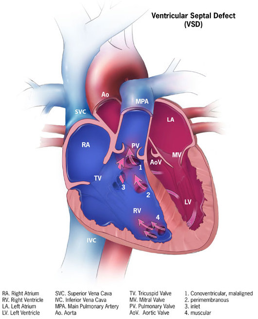
Types of VSD. Defects are classified by location, and location strongly predicts natural history, risk of complications, and treatment options (Baumgartner et al., European Heart Journal, 2021):
- Perimembranous VSDs are the most common type in clinical series (about 75–80%). They sit near the tricuspid and aortic valves and near the heart's electrical conduction system. That location matters for both the aortic valve and the risk of heart block during repair.
- Muscular VSDs are completely surrounded by muscle and are often multiple. They have the highest rate of spontaneous closure.
- Outlet VSDs (also called subarterial, supracristal, or doubly committed) sit just beneath the aortic and pulmonary valves. They rarely close on their own and can cause an aortic valve leaflet to prolapse into the defect, leading to progressive aortic regurgitation.
- Inlet VSDs (atrioventricular canal type) lie near the inflow valves. They are frequently associated with Down syndrome.
Physiology. The left ventricle pumps at much higher pressure than the right, so blood flows through the VSD from left to right. That extra blood circulates back through the lungs. The amount of flow depends on the size of the defect and on the resistance of the lung blood vessels.
Why symptoms often appear weeks after birth. At birth, resistance in the lung vessels is high, which limits flow through the defect. Over the first weeks of life, that resistance normally falls, and shunting increases. As a result, a large VSD often has no murmur or symptoms in the newborn nursery. Symptoms commonly emerge at about 4–8 weeks of age. This timeline is central to many VSD cases: a normal newborn exam and a normal pulse oximetry screen do not exclude a significant VSD. Because a VSD does not lower oxygen levels, pulse oximetry screening is not designed to detect it.
Natural History and Spontaneous Closure
Spontaneous closure is common, which is why observation is appropriate for many children:
- Copenhagen Baby Heart Study. This population-based study screened more than 25,000 newborns with echocardiography and found a VSD in 3.3%, mostly small muscular defects. By 1 year of age, 83.5% had closed. Muscular defects closed far more often than perimembranous defects (86.9% vs. 46.9%). Small size and a single defect predicted closure (Pihl et al., Neonatology, 2023).
- Chinese neonatal cohort. In a cohort followed for 7 years, 97.2% of muscular VSDs and 51.4% of perimembranous VSDs closed. A perimembranous defect of 4 mm or larger predicted that it would not close (Zhao et al., American Journal of Cardiology, 2019).
- Large defects. Large defects are much less likely to close, and the chance of closure falls steeply after the first 6–12 months of life.
Defects that stay open, even small ones, may later be associated with left heart enlargement, arrhythmia, aortic regurgitation, pulmonary hypertension, and endocarditis (Turner et al., Journal of the American College of Cardiology, 2022).
Clinical Presentation
Small (restrictive) VSD. These children have no symptoms and grow normally. A loud, harsh murmur is common. A loud murmur often means a small defect, because blood is forced at high velocity through a narrow opening.
Moderate to large VSD. These defects cause signs of congestive heart failure in infancy. According to the 2026 American College of Cardiology (ACC) guidance, these include fast heart rate, fast breathing, an enlarged liver, sweating, poor weight gain, and feeding intolerance (Sachdeva et al., Journal of the American College of Cardiology, 2026). Recurrent respiratory infections are also common.
Pitfalls in recognition:
- Heart failure symptoms in infants are easily attributed to reflux, formula intolerance, viral illness, or feeding technique.
- When a large VSD has caused high lung pressures, the murmur may become softer or disappear, because pressures in the two ventricles become nearly equal. A quieter murmur can mean the disease is getting worse, not better.
- Sweating during feeds and a downward trend on the growth chart are often the most objective early clues.
Eisenmenger syndrome. This is the end stage of an untreated large VSD. Years of excess lung blood flow can permanently damage the lung blood vessels. Resistance then rises above the body's resistance, the flow through the defect reverses from right to left, and the patient becomes cyanotic (blue) (Baumgartner et al., European Heart Journal, 2021). At that stage, closing the defect is generally no longer an option.
Diagnostic Evaluation
- Physical examination. Examination includes the murmur, breathing rate, liver size, and growth parameters.
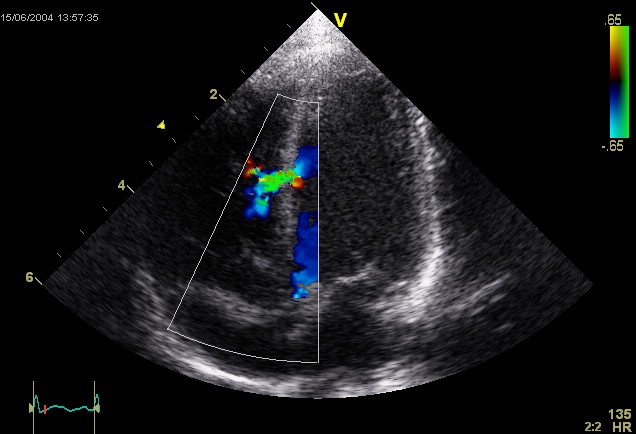
- Echocardiography. This is the definitive test. It shows the defect's location, size, and number; the direction and pressure gradient of flow; chamber enlargement; estimated lung pressures; aortic valve prolapse or regurgitation; and associated defects.
- Chest radiograph and ECG. These are supportive tests. They may show heart enlargement, increased lung blood flow, or ventricular hypertrophy, but they cannot exclude a VSD.
- Cardiac catheterization. This may be considered before intervention to measure lung vascular resistance, depending on the child's age and presentation (Sachdeva et al., Journal of the American College of Cardiology, 2026). It is usually unnecessary in young infants with heart failure and normal oxygen levels (Hansmann, Journal of the American College of Cardiology, 2017).
- Genetic evaluation. This is appropriate when the child has syndromic features. Down syndrome in particular changes the timing of surgery (see below).
Prenatal diagnosis. Large or malaligned VSDs may be seen on a fetal ultrasound. Small perimembranous and muscular defects are often not visible prenatally. A normal anatomy scan therefore does not exclude a VSD, and a missed small VSD seldom has clinical consequences.
Current Evidence-Based Management
Observation. Small defects in children without symptoms and without left heart enlargement are typically followed with periodic echocardiography to watch for closure or complications.
Medical therapy. The 2026 ACC guidance recommends starting medical therapy in symptomatic patients. Options include diuretics, afterload-reducing medications, calorie-dense feeds, and nasogastric (tube) feeding when needed (Sachdeva et al., Journal of the American College of Cardiology, 2026).
Referral for closure. According to the 2026 ACC guidance:
- Closure, surgical or catheter-based, is indicated if medical therapy does not improve symptoms or left heart enlargement, or if other complications appear.
- Closure of large defects should be considered during infancy even without symptoms, because of the risk of irreversible pulmonary vascular changes (Sachdeva et al., Journal of the American College of Cardiology, 2026).
Timing. Commonly cited timing principles include:
- Failure to thrive from heart failure that does not respond to medical management is a widely accepted indication for closure in infants younger than 6 months. Significant pulmonary hypertension is an accepted indication in infants older than 6 months (Hekim Yilmaz et al., Catheterization and Cardiovascular Interventions, 2025).
- Pulmonary hypertension experts describe repair of a VSD within the first 2 years of life as essential to avoid irreversible lung vascular changes. In children with added risk, such as Down syndrome, closure within the first 6 months is advised (Hansmann, Journal of the American College of Cardiology, 2017).
Other indications for closure:
- Prolapse of an aortic valve leaflet with progressive aortic regurgitation can be an indication for VSD closure in children, to avoid early aortic valve surgery.
- A prior episode of endocarditis in a child with a small VSD may prompt consideration of closure.
These indications appear in the 2025 ACC/AHA adult congenital heart disease guideline (Gurvitz et al., Journal of the American College of Cardiology, 2026).
Surgical vs. transcatheter closure.
- Surgical patch closure remains the standard for most defects. It is generally low risk with good long-term results.
- Catheter-based device closure works well for many muscular defects.
- Perimembranous defects carry a notable risk of complete heart block with device closure. No FDA-approved device for perimembranous VSD is available in the United States (Gurvitz et al., Journal of the American College of Cardiology, 2026).
Guidelines vs. legal standards. Guideline recommendations describe evidence-based best practice. They are not the same as the legal standard of care, which is set by jurisdiction and judged against what a reasonable practitioner would have done in similar circumstances at that time.
Complications of VSD and Its Treatment
Disease-related complications:
- Heart failure and failure to thrive from large shunts.
- Pulmonary arterial hypertension, progressing to Eisenmenger syndrome. This is the most consequential complication and the one most tied to timing.
- Aortic valve prolapse and aortic regurgitation, mainly with outlet and some perimembranous defects. A sinus of Valsalva aneurysm (a bulge in the aortic root) may also form and can rupture (Gurvitz et al., Journal of the American College of Cardiology, 2026).
- Double-chambered right ventricle and subaortic membrane, which are obstructions that can develop over time.
- Infective endocarditis. This is not uncommon in patients with unrepaired VSDs or residual defects and can have devastating effects. In Eisenmenger physiology, endocarditis carries a risk of stroke and brain abscess (Gurvitz et al., Journal of the American College of Cardiology, 2026).
- Arrhythmias, particularly later in life.
Procedure-related complications:
- Complete heart block. The conduction system runs along the edge of perimembranous defects. After device closure of perimembranous VSDs, heart block has been reported in 2–22% of cases. It can appear immediately or weeks to months later and may be transient or permanent, requiring a pacemaker (Turner et al., Journal of the American College of Cardiology, 2022). Surgical closure also carries a smaller, well-recognized risk.
- Residual shunt, valve injury (tricuspid or aortic), device embolization, hemolysis, and vascular injury. Patients weighing under 5 kg have a higher risk of device-related complications (Turner et al., Journal of the American College of Cardiology, 2022).
- Long-term sequelae. A cohort followed up to 40 years after surgical closure found that most patients had good functional status. However, a meaningful minority had arrhythmias, needed reintervention, or developed ventricular dysfunction or aortic regurgitation. The authors argued for continued surveillance rather than discharge from cardiology care (Menting et al., Journal of the American College of Cardiology, 2015).
Medical Considerations in Evaluating Standards of Care
Expert reviewers commonly assess the following:
- Murmur evaluation. Was a new or changing murmur appropriately characterized? Was it reassessed? Was it referred when its features, or the child's symptoms, were not consistent with an innocent murmur?
- Recognition of heart failure. Were feeding difficulty, sweating, fast breathing, and falling growth percentiles recognized as possible cardiac symptoms rather than attributed only to feeding or gastrointestinal causes?
- Growth monitoring. Were growth curves plotted and acted on?
- Referral timeliness. Once a significant VSD was suspected, was cardiology referral and echocardiography arranged with urgency matched to the clinical picture?
- Cardiology surveillance. Were follow-up intervals appropriate for the type and size of the defect? Were aortic valve prolapse, worsening regurgitation, left heart enlargement, or rising lung pressures identified and acted on?
- Timing of intervention. Was closure considered within the accepted window, especially for large defects and for children with Down syndrome?
- Procedural decisions. Was the choice between surgery and a device, and the selection of patients, reasonable? Were risks such as heart block disclosed? Was rhythm monitoring after the procedure adequate, given that heart block can appear late?
- Informed consent. Did the consent discussion cover heart block, possible pacemaker need, residual defect, valve injury, and the alternatives?
- Communication and documentation. Were parental concerns documented and addressed? Were follow-up instructions clear? Were handoffs between primary care and cardiology complete?
Hindsight bias is a constant risk. Feeding problems and murmurs are extremely common in infancy, and most are benign. The question is whether the information available at each encounter reasonably called for a different response.
Common Allegations in Litigation
- Failure to evaluate a murmur. In a child with a pathologic murmur or symptoms, the murmur was labeled innocent without reassessment or referral.
- Failure to recognize heart failure or failure to thrive. Symptoms were attributed to reflux, formula intolerance, or parental anxiety.
- Delayed cardiology referral or echocardiography.
- Missed or delayed closure, resulting in irreversible pulmonary vascular disease or Eisenmenger syndrome.
- Failure to monitor for aortic valve prolapse, leading to valve damage that needs repair or replacement.
- Procedural injury, including heart block requiring a pacemaker, residual shunt, valve injury, or device embolization.
- Inadequate informed consent for device closure, particularly for perimembranous defects.
- Loss to follow-up of a child known to have a VSD, through system or communication failures.
Each allegation has a balancing medical context. Most murmurs are innocent. Feeding problems are nearly universal in infancy. Many VSDs close on their own, so watchful waiting is often appropriate. Some procedural complications occur despite proper technique.
Medical Causation
Causation analysis in VSD cases usually turns on a few questions:
- Pulmonary vascular disease. Would earlier referral and closure, within the accepted window, more likely than not have prevented persistent pulmonary hypertension? Answering this requires reconstructing when lung vascular changes became established. Factors include age at repair, catheterization data, genetic susceptibility such as Down syndrome, and associated lesions.
- Growth and neurodevelopment. Poor growth and illness from untreated heart failure may contribute to developmental concerns. However, prematurity, genetic syndromes, and the risks of surgery itself are confounders.
- Aortic valve injury. Would earlier closure have prevented progression to the need for valve surgery? Evidence on rates of progression in adults is limited (Gurvitz et al., Journal of the American College of Cardiology, 2026).
- Procedural complications. Did the injury result from a deviation, such as improper device sizing, poor patient selection, or delayed recognition of heart block? Or was it a known risk that can occur even with proper technique?
- Probability versus possibility. Opinions generally must be held to a reasonable degree of medical probability (more likely than not). Showing that an earlier intervention might possibly have helped is generally not sufficient.
Long-Term Outcomes
- Children with small VSDs that close, and those with timely, uncomplicated repair, generally do very well.
- Survival decades after surgical closure appears slightly lower than in the general population. Late arrhythmias, reinterventions, and aortic regurgitation occur in a minority of patients (Baumgartner et al., European Heart Journal, 2021; Menting et al., Journal of the American College of Cardiology, 2015).
- Patients with Eisenmenger syndrome face markedly reduced life expectancy, limits on exercise, risks of bleeding and clotting, and high-risk pregnancy. Pregnancy is generally discouraged in this group.
- Patients who need a pacemaker after closure require lifelong device management and generator replacements.
- Lifelong congenital cardiology follow-up is advised for patients with residual defects, pulmonary hypertension, valve disease, or conduction abnormalities.
Medical Records That Often Matter
- Prenatal ultrasound and any fetal echocardiogram.
- Nursery records, including the newborn exam and pulse oximetry screen. A normal screen is expected with a VSD.
- Well-child visit records, including murmur descriptions over time, growth charts, feeding history, and documented parental concerns.
- Urgent care and emergency department records.
- Echocardiogram reports and images, including defect type and size, gradients, chamber size, estimated lung pressures, and aortic valve assessment.
- Cardiology notes, including recommended follow-up intervals and whether visits actually occurred.
- Catheterization data, including measured lung vascular resistance.
- Operative and catheterization procedure reports, consent forms, and device details.
- Postoperative ECGs and rhythm monitoring.
- Long-term follow-up records, including pulmonary hypertension care, pacemaker checks, and developmental assessments.
Common Misconceptions
- "A loud murmur means a big hole." Often the opposite is true. Small defects are frequently the loudest.
- "A normal newborn exam and pulse oximetry screen exclude a VSD." They do not. Murmurs and symptoms commonly appear only after lung resistance falls in the first weeks of life, and pulse oximetry is not designed to detect VSDs.
- "A murmur that gets quieter means the hole is closing." It may instead mean that lung pressures are rising.
- "Every VSD needs surgery." Most small defects, especially muscular ones, close on their own (Pihl et al., Neonatology, 2023).
- "Heart block after closure always means a technical error." Heart block is a recognized risk, particularly with perimembranous defects. Its cause must be assessed case by case.
- "Once repaired, the patient is cured." Late complications can occur decades later (Menting et al., Journal of the American College of Cardiology, 2015).
The Value of Pediatric Cardiology Expertise
VSD cases sit where general pediatrics and pediatric cardiology meet. A reviewer needs to be able to judge:
- what a reasonable pediatrician should recognize at a routine visit;
- how defect type and size predict natural history;
- echocardiographic and catheterization data;
- the timing of intervention relative to pulmonary vascular risk;
- the risks of surgical versus catheter-based closure;
- the long-term outcome literature needed for causation and damages analysis.
Expertise in both general pediatrics and congenital cardiology allows each phase of care to be evaluated objectively.
Key Takeaways for Attorneys
- VSD is the most common congenital heart defect. Most small defects, especially muscular ones, close spontaneously within the first year.
- Defect location (perimembranous, muscular, outlet, inlet) strongly predicts natural history, complications, and procedural risk.
- Large VSDs often cause no symptoms in the nursery. Heart failure typically appears at about 4–8 weeks, as lung resistance falls.
- A normal newborn exam and pulse oximetry screen do not exclude a significant VSD.
- Feeding difficulty, sweating, fast breathing, and falling growth percentiles are key early signs of heart failure.
- Large defects that do not respond to medical therapy, or that cause pulmonary hypertension, should be considered for closure in infancy. Earlier closure is advised in Down syndrome.
- Irreversible pulmonary vascular disease (Eisenmenger syndrome) is the most consequential timing-dependent outcome.
- Complete heart block, residual shunt, and valve injury are recognized procedural risks. Their occurrence alone does not establish negligence.
- Causation requires individualized analysis to a reasonable degree of medical probability, accounting for genetics, defect anatomy, and the natural history of the disease.
- Objective review by physicians experienced in both general pediatrics and pediatric cardiology is essential to separate preventable harm from inherent risk.
This article is for educational purposes only. It does not constitute legal advice or an opinion regarding any specific case.
References
- Baumgartner et al. European Heart Journal. 2021.
- Pihl et al. Neonatology. 2023.
- Zhao et al. American Journal of Cardiology. 2019.
- Turner et al. Journal of the American College of Cardiology. 2022.
- Sachdeva et al. Journal of the American College of Cardiology. 2026.
- Hansmann. Journal of the American College of Cardiology. 2017.
- Hekim Yilmaz et al. Catheterization and Cardiovascular Interventions. 2025.
- Gurvitz et al. Journal of the American College of Cardiology. 2026.
- Menting et al. Journal of the American College of Cardiology. 2015.
Educational Disclaimer
This article is provided for educational purposes only and does not constitute legal or medical advice. Every case is unique and should be evaluated on its individual facts and medical records. Reading this article does not create an expert-client relationship.
About the Author
Asif Masood, MD, MSc is double board-certified in General Pediatrics and Pediatric Cardiology. Expert witness services in general pediatrics and pediatric & congenital cardiology, complemented by independent, non-testimony medical consulting across a broad range of medical specialties. Through The Verdict MD, he serves attorneys, law firms, insurers, and healthcare organizations nationwide.
Learn about Dr. Masood's pediatric cardiology expert witness services for independent review of the medical questions in your case.